. 2004 Mar 31;24(13):3402-12.
doi: 10.1523/JNEUROSCI.5241-03.2004.

Beta1-integrins are critical for cerebellar granule cell precursor proliferation

Affiliations

Beta1-integrins are critical for cerebellar granule cell precursor proliferation

Sandra Blaess et al. J Neurosci. .

Abstract

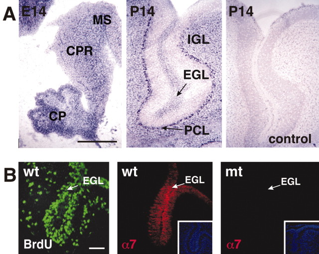
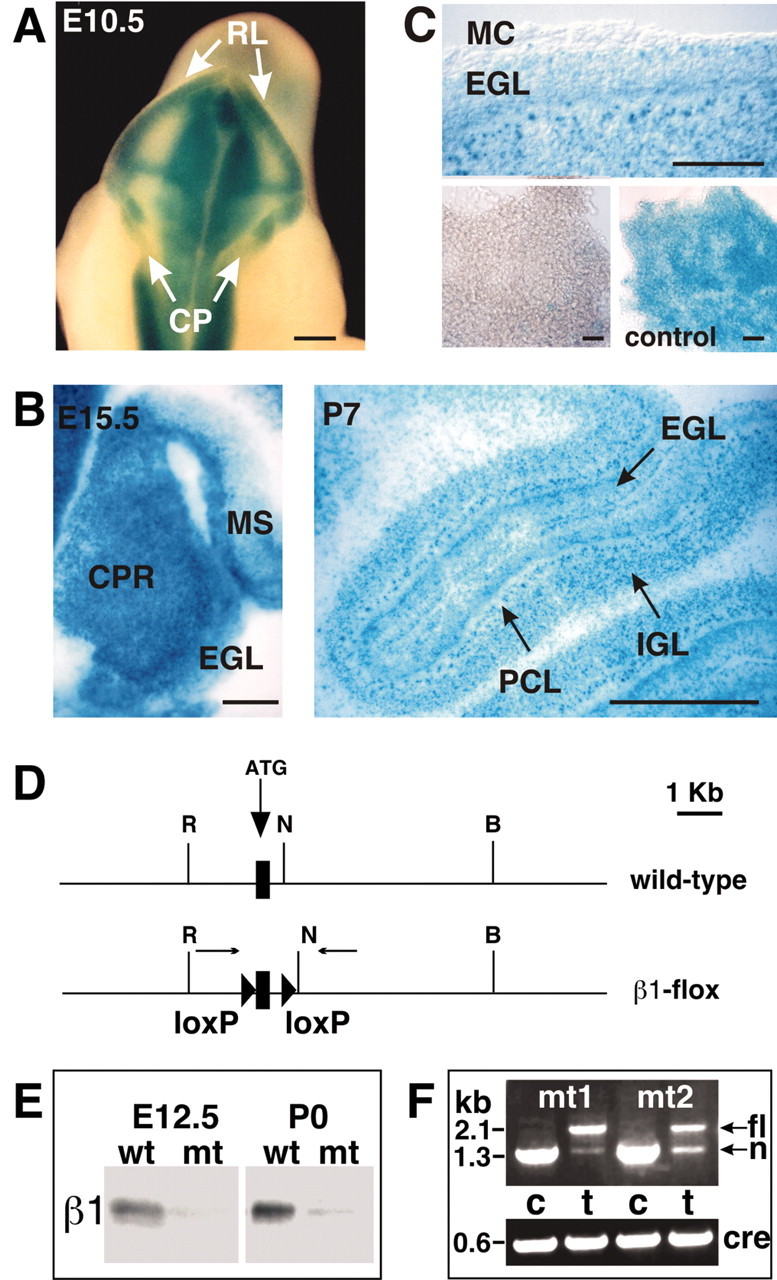
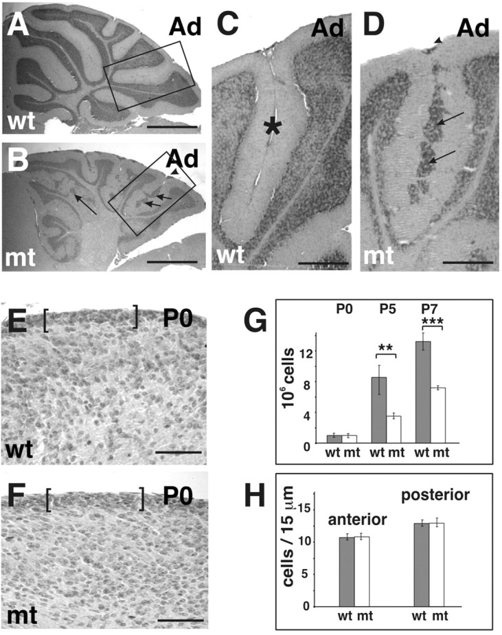
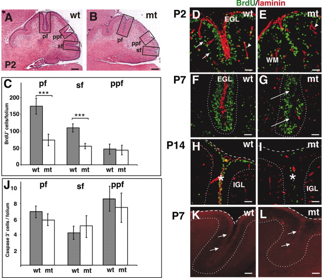
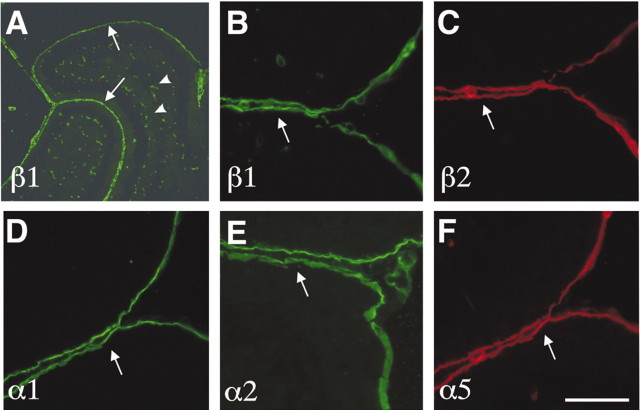
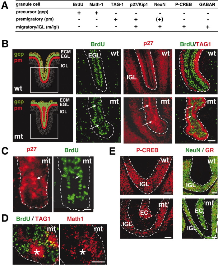
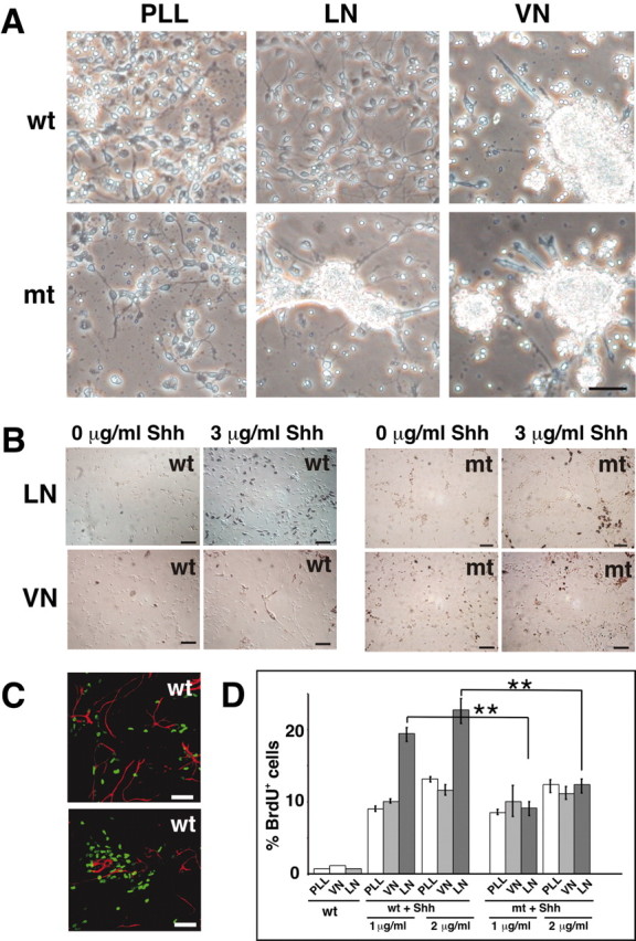
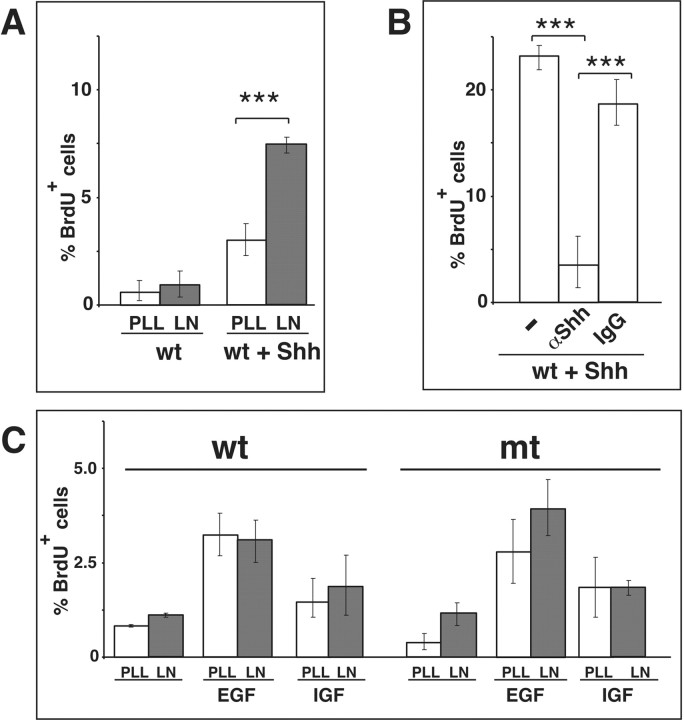
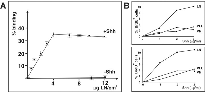
We have previously shown that mice with a CNS restricted knock-out of the integrin beta1 subunit gene (Itgb1-CNSko mice) have defects in the formation of lamina and folia in the cerebral and cerebellar cortices that are caused by disruption of the cortical marginal zones. Cortical structures in postnatal and adult Itgb1-CNSko animals are also reduced in size, but the mechanism that causes the size defect has remained unclear. We now demonstrate that proliferation of granule cell precursors (GCPs) is severely affected in the developing cerebellum of Itgb1-CNSko mice. In the absence of beta1 expression, GCPs lose contact with laminin in the meningeal basement membrane, cease proliferating, and differentiate prematurely. In vitro studies provide evidence that beta1 integrins act at least in part cell autonomously in GCPs to regulate their proliferation. Previous studies have shown that sonic hedgehog (Shh)-induced GCP proliferation is potentiated by the integrin ligand laminin. We show that Shh directly binds to laminin and that laminin-Shh induced cell proliferation is dependent on beta1 integrin expression in GCPs. Taken together, these data are consistent with a model in which beta1 integrin expression in GCPs is required to recruit a laminin-Shh complex to the surface of GCPs and to subsequently modulate the activity of signaling pathways that regulate proliferation.


Figures

Figure 1.
Figure 1.
Integrin expression in the cerebellum. A, In situ hybridization was performed with an Itgb1-specific probe on sagittal sections. In wild-type mice, Itgb1 was expressed throughout the cerebellar primordium (CPR), choroid plexus (CP), and mesencephalon (MS) at E14, and in the external granule cell layer (EGL), the internal granule cell layer (IGL), and the Purkinje cell layer (PCL) at P14. No signal was detected with the sense control probe (control). B, Left panel, Proliferating GCPs in the EGL were visualized by staining sections of mice injected with BrdU with antibodies to BrdU. Middle panel, Immunostaining for the integrin α7 subunit (red). Sagittal sections from a P2 wild-type cerebellum revealed thatα7 expression was restricted to the EGL. A single folium is shown. Inset panel shows the same areas stained with DAPI to reveal nuclei. Left panel, Expression of the integrin α7 subunit was not detectable in the EGL of Itgb1-CNSko mice. Scale bars: A, 200 μm; B, 50 μm.
Figure 2.
Figure 2.
Cre recombination pattern and inactivation of the Itgb1 gene in the cerebellum. A–C, Offspring of intercrosses between Rosa26lacZflox mice and nestin-Cre mice were analyzed for LacZ expression to determine the Cre recombination pattern in the cerebellum. A, At E10.5, recombination was evident in whole-mount staining in the rhombic lip (RL), but not in the developing choroid plexus (CP). B, In histological sections of E15.5 embryos, recombination was apparent throughout the cerebellar primordium (CPR) including the forming EGL. Recombination also occurred in the mesencephalon (MS). At P7, recombination was evident in all cell layers of the cerebellum, including the EGL, IGL, and PCL. Note that the P7 sections were cut thinner than the E15.5 sections, revealing the nuclear localization of LacZ. C, The top panel shows a detail of a sagittal P7 section, revealing that the meningeal cell layer (MC) was not recombined. The bottom panels show meningeal cell layers dissected from P7 mice. Meninges were spread out on a coverslip and photographed. Nestin-Cre did not induce recombination in the meninges (bottom left panel). In control mice that carried a fully recombined Rosa26lacZflox locus, the meninges were LacZ-positive (bottom right panel, control). D, Diagram of the Itgb1 wild-type and Itgb1flox alleles. The first coding exon (rectangle), the lox P sites (triangles), the primers used for analyzing Cre-mediated recombination (arrows), and several restriction endonuclease cleavage sites are indicated (R = EcoRI; N = NheI; B = BamHI). E, The neural tube from E12.5 mice and the cerebellar anlage from P0 mice were dissected. Protein extracts were analyzed by Western blotting. β1 protein was expressed in wild-type mice, but very low amounts in the mutants. Low amounts of β1 protein in the mutants were expected because of β1 expression in blood vessels and meninges. F, DNA was prepared from the cerebellum (c) and tail (t) of two mutant mice at P0 and analyzed by PCR for Cre-mediated recombination. In mouse tail DNA the 2.1 kb Itgb1flox allele (fl) and the 1.3 kb Itgb1null allele (n) were detectable. In the cerebellar DNA only a 1.3 kb band indicative of both the Itgb1null allele and the recombined Itgb1flox allele was apparent. Primers against Cre were used to confirm the presence of the Cre transgene. Scale bars, 200 μm.
Figure 3.
Figure 3.
Cerebellar defects in Itgb1- CNSko mice. A, B, Sagittal sections of adult (Ad) animals were stained with hematoxylin–eosin. The cerebellar cortex of wild-type (wt) mice showed a regular foliation pattern. The cerebellar cortex of the mutant (mt) mice lacked fissures and the depth of folia was reduced, but a rudimentary internal foliation pattern was discernible from the shape of the IGL. Note that the overall thickness of the IGL was drastically reduced. Granule cell ectopia remained along the fusion lines of folia (arrows) and on the surface of the cerebellum (arrowhead). C, D, Higher magnification view of the areas indicated in A and B. In wild-type mice, adjacent cerebellar folia were separated by fissures (asterisks). In mutants, adjacent folia fused and granule cell ectopia formed at the cerebellar surface (arrowhead) and along the fusion line (arrows). E, F, Part of a midsagittal sections through the central lobe of the cerebellum of a wild-type and mutant mouse at P0. The EGL is indicated by brackets. G, The cerebellum was dissected from animals of the indicated age and dissociated. The total cell number was determined. For each age, cell counts were performed for three wild-type and three mutant animals. Error bars indicate the SD. A Student's t test was performed (***p < 0.001; **p < 0.01). Scale bars: A, B, 600 μm; C, D, 100 μm; F, G, 50 μm. H, Cell numbers in the EGL were determined both in the anterior and posterior aspect of the central lobe. Cells were counted on two adjacent midsagittal sections for two wild-type and two mutant mice. The mean and SD were determined.
Figure 4.
Figure 4.
Defective granule cell precursor proliferation. A, B, Sagittal sections of P2 animals were stained with hematoxylin–eosin. The folia in the mutant mice lacked fissures, and adjacent EGLs within the folia were fused. Note that in the mutants the EGL in some folia was thinner than in the wild-type mice. Boxed areas indicate folia that would be separated by the primary fissure (pf), prepyramidal fissure (ppf), and secondary fissure (sf). C–I, Mice were injected with BrdU and killed 1 hr later. Sagittal cerebellar sections were analyzed for BrdU incorporation. C, The number of proliferating GCPs was determined at P2 by counting BrdU-positive cells in the EGL of the folia indicated in A and B. Two sections were counted per animal (2 wild-type and 2 mutant mice). Error bars indicate the SD. A Student's t test was performed (***p < 0.001). D–I, Double staining for BrdU (green) and LN (red). D, E, Example of a P2 folium. At P2, proliferation was greatly diminished in the mutant cerebellum, in particular within the developing folia. Note that LN was incorporated into the folium in the wild-type cerebellum but was absent in the mutant. Scattered BrdU-positive cells in deeper layers of the cerebellum (arrows) were probably glial cells (WM, white matter). Arrowheads indicate LN immunostaining around blood vessels. F, G, At P7, BrdU-negative ectopia formed in the fused folia (arrows) in the mutant, in areas where GCPs in wild-type mice were in contact with ECM components. The EGL–PCL boundary is outlined. H, I, BrdU labeling at P14 revealed strong proliferative defects at the cerebellar surface (dashed line), and almost complete absence of proliferation within the folia (asterisks) of the mutants. The IGL–PC border is outlined (dotted line). At this stage the basement membrane component LN was also absent from the cerebellar surface (dashed line) in the mutant. J, The number of apoptotic cells was determined at P7 by counting caspase 3-positive cells in the EGL of the folia indicated in A and B. Five to seven sections were counted each in three wild-type and two mutant mice. Error bars indicate the SD. K, L, Caspase 3 staining (red) at P7. The PCL–IGL boundary (dotted line) and the cerebellar surface (dashed line) is outlined. Arrows point to caspase 3-positive cells. Scale bars: A, B, 100 μm; D–I, 40 μm; K, L, 60 μm.
Figure 5.
Figure 5.
Expression of LN subunits in the cerebellum. Sagittal cerebellar sections were stained with antibodies directed against different LN subunits as indicated in the panels. A, Part of a cerebellum at P10 is shown. B–F, High-magnification view of cerebellar folia at P7. The arrows point to the meningeal basement membranes adjacent to the EGL. The arrowheads point to blood vessels. Scale bars: A, 400 μm; B–F, 50 μm.
Figure 6.
Figure 6.
Premature differentiation of granule cells. The expression of molecular markers was analyzed to determine the differentiation state of BrdU-negative granule cells within the proliferation zone of the EGL. A, Summary of the expression patterns of molecular markers during granule cell differentiation [gcp, GCPs in the EGL; pm, premigratory granule cells in the EGL; m/igl, migrating cells and cells in the IGL; (–), no expression; (+), expression]. B–D, Analysis of P7 wild-type and mutant cerebella (sagittal sections). Mice were injected with BrdU and killed 1 hr later. B, The left panels show a schematic illustration of the differentiation states of granule cells in the EGL of a wild-type (top panel) and mutant (bottom panel) folium at P7. The boxed area indicates the region shown in the subsequent immunostainings. Immunostaining for BrdU (green) and p27–Kip1 (red) on adjacent sections: in wild-type, postmitotic but not BrdU-positive cells expressed p27–Kip1. In the mutant, BrdU-negative ectopia formed in the fused folia (arrows) in areas in which GCPs in wild-type mice are in contact with ECM components (Fig. 4 D,E). p27–Kip1 was expressed in the BrdU-negative ectopia in the mutants (arrows). Double staining for BrdU (green) and TAG-1 (red): GCPs that ceased to proliferate started to differentiate in ectopic locations (arrows). C, D, Higher magnification of a tip of a folium in the mutant. C, Staining for p27–Kip1 (red) and BrdU (green): granule cells that ceased to proliferate upregulated p27–Kip1 (arrow). D, Double staining for BrdU (green) and TAG-1 (red) or Math-1 (red) on an adjacent section at P7: the BrdU-negative ectopia expressed TAG-1, but not Math-1 (asterisk). Thus, the cells lost their GCP character in an ectopic location and initiated differentiation. The EGL is outlined. E, Staining for P-CREB (red), or NeuN (green) and GABARα6 (GR; red) in sections from P14 animals: in wild-type mice P-CREB, NeuN, and GABARα6 were expressed only in the IGL. In contrast, these proteins were expressed in mutant mice in nonproliferating cells in ectopic positions (EC) in the EGL. Scale bars, 40 μm.
Figure 7.
Figure 7.
Effects of Shh and ECM on granule cell precursor proliferation. A, Wild-type and Itgb1-deficient cells were plated in plastic tissue culture wells coated with PLL, LN-1, or VN. Phase-contrast images of cells after 1 d in culture. Note that wild-type granule cells adhered to and spread on PLL and LN-1, but not on VN, and that adhesion to LN-1 was abolished in Itgb1-deficient cells. B, Cerebellar cells were plated on PLL–LN-1 or PLL–VN and cultured for 3 d in the absence or presence of Shh. Before fixation, cultures were pulsed with BrdU for 4 hr and stained with an anti-BrdU antibody (brown color). Both wild-type (wt) and Itgb1-deficient (mt) cells were plated and compared. C, Immunostaining for GFAP (red) and BrdU (green) showing that the proliferating cells are not GFAP-positive glial cells. D, Quantification of BrdU-positive wild-type and mutant cells cultured in the presence or absence of Shh (1 and 2 μg/ml) on ECM substrates. Analysis was performed by counting total cells and BrdU-immunostained cells. BrdU-positive cells were expressed as a percentage of the total cell number. A minimum of 1000 cells was counted per experiment. A Student's t test was performed (**p ≤ 0.01). Scale bars, 40 μm.
Figure 8.
Figure 8.
β1 integrins and LN-1 cooperate with Shh but not with EGF or IGF to regulate GCP proliferation. A, Granule cells were purified by Percoll gradient centrifugation, plated on the indicated substrates and, where indicated, were treated with 1 μg/ml Shh. Quantification was performed in three independent experiments as described in Figure 7D. The mean and SD were determined, and a Student's t test was performed (***p < 0.001). B, Cells were cultured on LN-1 in the presence of 1 μg/ml Shh. Either no antibody (–) was added, 50 μg/ml antibody to Shh (α-Shh), or 50μg/ml control IgG. The quantification was performed as described in Figure 7D. C, Cells were cultured on PLL or PLL/LN-1 for 3 d in the presence of 50 ng/ml EGF or IGF. The cells were labeled for the final 6 hr of culturing with BrdU, and the percentage of BrdU-positive cells was determined. The results show the mean and SD from three different experiments.
Figure 9.
Figure 9.
Shh binds to LN-1. A, Increasing amounts of LN-1 were coated on 96 well plates, blocked with BSA, and 1 μg/ml Shh was added (+Shh) or omitted (–Shh). The plates were washed with PBS, and bound Shh was detected by ELISA assays. The values were normalized against 1 μg/ml Shh that was directly bound to plastic (100%). The experiment was performed three times, and the mean and SD were determined. B, Dishes were precoated with ECM substrates, incubated with the indicated amounts of Shh, and subsequently washed. Wild-type cells were plated onto the substrates and cultured for 3 d without further addition of Shh. The cells were labeled for the final 4 hr of culturing with BrdU, and the number of BrdU-positive cells was quantified as described in the legend to Figure 7. Two independent representative experiments are shown.

Similar articles

Cited by

References

    1. Altman J, Bayer SA (1997) Development of the cerebellar system in relation to its evolution, structure, and function. New York: CRC.
    1. Baeg GH, Perrimon N (2000) Functional binding of secreted molecules to heparan sulfate proteoglycans in Drosophila Curr Opin Cell Biol 12: 575–580. - PubMed
    1. Barakat I, Wittendorp-Rechenmann W, Rechenmann RV, Sensenbrenner M (1981) Influence of meningeal cells on the proliferation of neuroblasts in culture. Dev Neurosci 4: 363–372. - PubMed
    1. Bellaiche Y, The I, Perrimon N (1998) Tout-velu is a Drosophila homologue of the putative tumour suppressor EXT-1 and is needed for Hh diffusion. Nature 394: 85–88. - PubMed
    1. Bhatt RS, Tomoda T, Fang Y, Hatten ME (2000) Discoidin domain receptor 1 functions in axon extension of cerebellar granule neurons. Genes Dev 14: 2216–2228. - PMC - PubMed

Publication types

MeSH terms