. 2004 Apr 21;23(8):1739-48.
doi: 10.1038/sj.emboj.7600136. Epub 2004 Apr 1.

E-cadherin-mediated adhesion inhibits ligand-dependent activation of diverse receptor tyrosine kinases

Affiliations

E-cadherin-mediated adhesion inhibits ligand-dependent activation of diverse receptor tyrosine kinases

Xiaolan Qian et al. EMBO J. .

Abstract

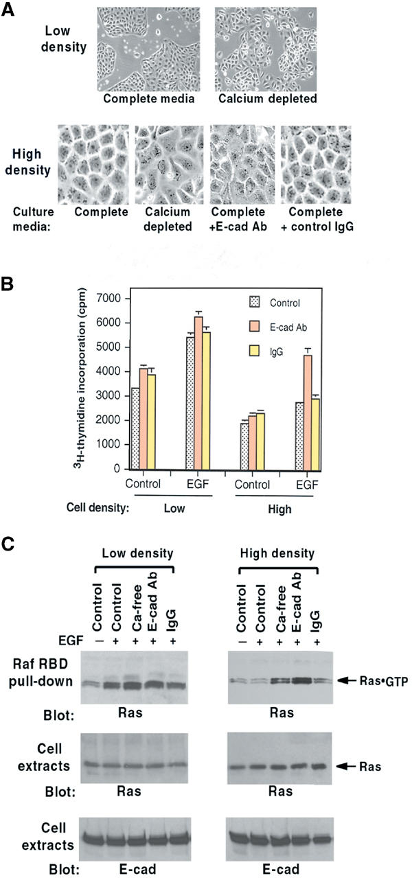
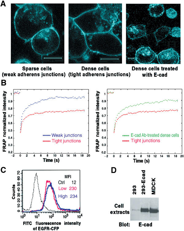
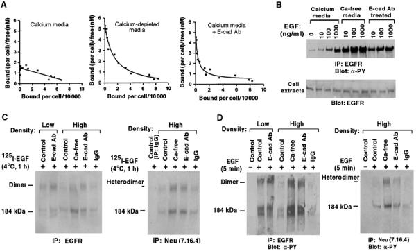
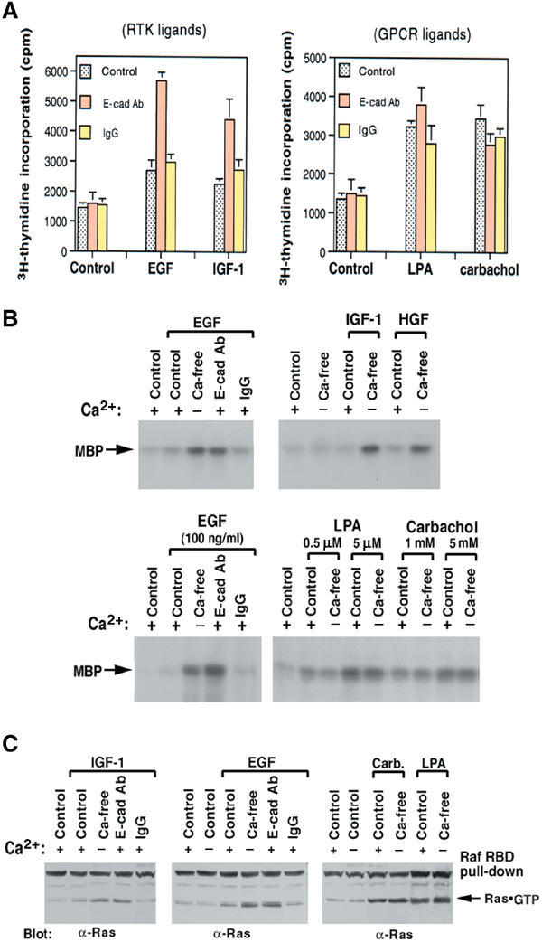
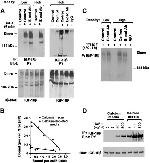
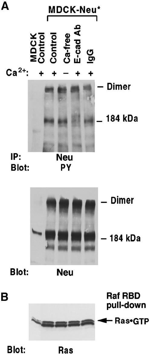
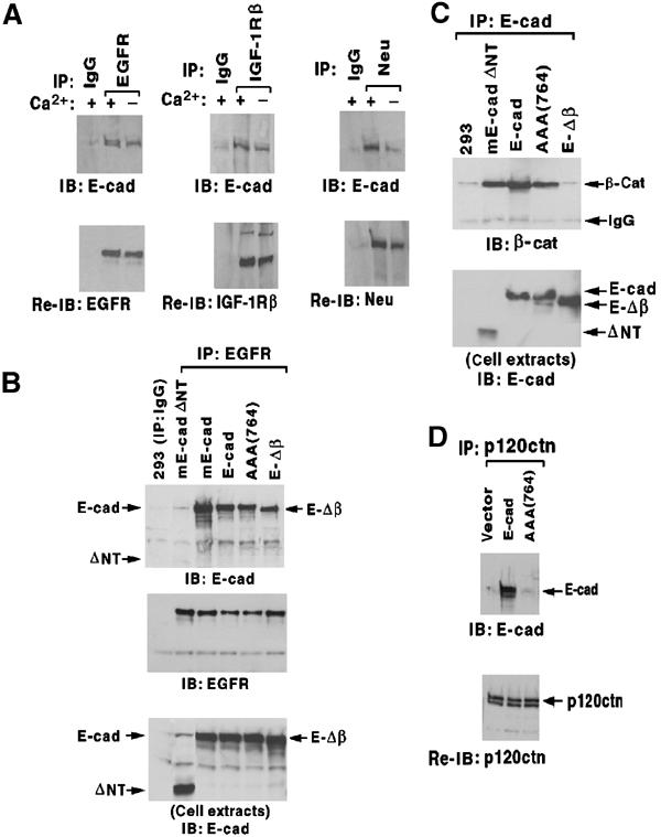
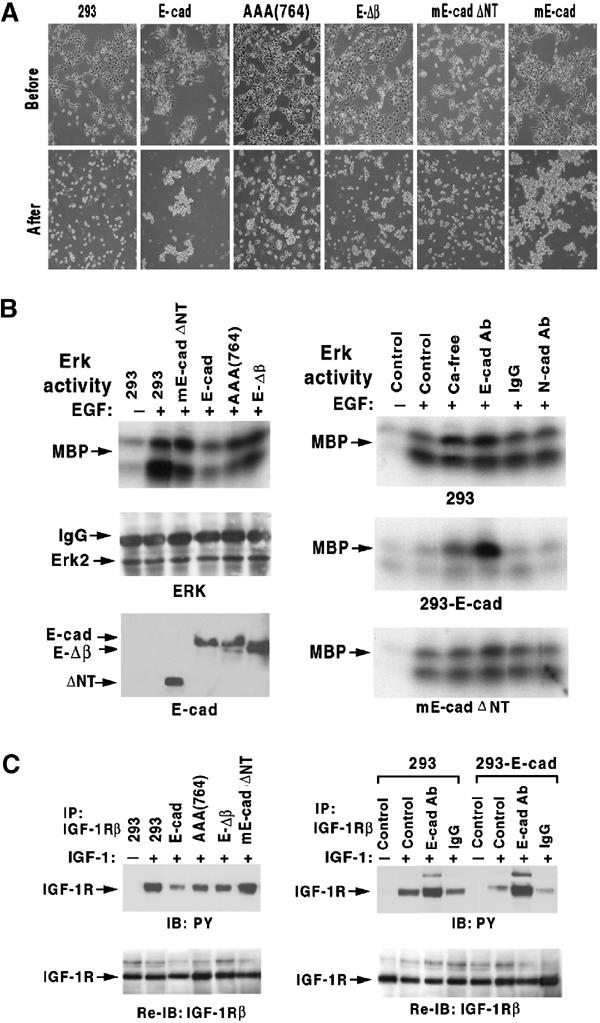
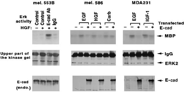
E-cadherin is an essential adhesion protein as well as a tumor suppressor that is silenced in many cancers. Its adhesion-dependent regulation of signaling has not been elucidated. We report that E-cadherin can negatively regulate, in an adhesion-dependent manner, the ligand-dependent activation of divergent classes of receptor tyrosine kinases (RTKs), by inhibiting their ligand-dependent activation in association with decreases in receptor mobility and in ligand-binding affinity. E-cadherin did not regulate a constitutively active mutant RTK (Neu*) or the ligand-dependent activation of LPA receptors or muscarinic receptors, which are two classes of G protein-coupled receptors. EGFR regulation by E-cadherin was associated with complex formation between EGFR and E-cadherin that depended on the extracellular domain of E-cadherin but was independent of beta-catenin binding or p120-catenin binding. Transfection of E-cadherin conferred negative RTK regulation to human melanoma and breast cancer lines with downregulated endogenous E-cadherin. Abrogation of E-cadherin regulation may contribute to the frequent ligand-dependent activation of RTK in tumors.


Figures

Figure 1
Figure 1
Effects of MDCK cell density on EGF responses. (A) Morphology of MDCK cells at low and high density and effects of 16 h incubation with calcium depletion, E-cadherin neutralizing antibody (E-cad Ab), or control IgG. Original magnifications were the same; images on the top panels are × 4, and those on the bottom panels × 10. (B) DNA synthesis in cells treated as in (A) and then given EGF (10 ng/ml) for 24 h. (C) Ras·GTP response of cells treated as in (A) and then stimulated with EGF (100 ng/ml) for 5 min. Ras·GTP was determined by Raf RBD pull-down in the upper panel. Total Ras and E-cadherin in cell extracts are in the middle and lower panels, respectively.
Figure 2
Figure 2
Adhesion-dependent inhibition of EGFR-CFP mobility. (A) Confocal images of HEK293 cells that stably express E-cadherin (293-Ecad) were transiently transfected with a plasmid encoding EGFR-CFP. Right panel: Cells treated with E-cadherin neutralizing antibody for 16 h. Original magnification × 100. Each bar represents 10 μm. (B) Mean FRAP values of EGFR-CFP in cells as in (A). Cell–cell junction regions from sparse, dense, or E-cad Ab-treated dense cells (as shown in panel A) were subjected to FRAP analysis. The normalized mean curves from weak versus tight adherens junctions were compared (left panel); the tight junction curve was also compared with that from the E-cad Ab-treated cells (right panel). The standard errors were calculated for each time point, but are shown only for the last time point. (C) FACS analysis of EGFR-CFP expression on the surface of cells as in (A). Cells were trypsinized and stained with anti-EGFR mAb 225 and FITC-conjugated anti-mouse IgG. MFI=mean fluorescence intensity. (D) E-cadherin expression in 293-Ecad cells. Equal amounts of cell extracts from parental HEK293, 293-Ecad, and MDCK cells were analyzed by anti-E-cadherin blotting.
Figure 3
Figure 3
EGF binding and activation of EGFR and wild-type Neu in MDCK cells. (A) Scatchard analysis of EGF binding to MDCK-EGFR cells. Confluent MDCK-EGFR cells pretreated with media as indicated were assayed for specific EGF binding. The results were analyzed by the Scatchard method to obtain dissociation constants Kd (nM) and number of binding sites per cell (see text). The plots represent the best fit for the data. (B) High concentrations of EGF can overcome the adhesion-dependent inhibition of EGFR activation. Confluent MDCK-EGFR cells were pretreated as in Figure 1 and stimulated with the indicated concentrations of EGF for 5 min. Cell extracts were immunoprecipitated with anti-EGFR followed by antiphosphotyrosine blots (upper panel) or directly analyzed with anti-EGFR blots as loading controls (lower panel). (C) Crosslinking of 125I-EGF to EGFR-EGFR homodimers and EGFR-Neu heterodimers. MDCK-EGFR cells were incubated with 0.4 nM of 125I-EGF for 1 h at 4°C followed by treatment with BS3, immunoprecipitated with EGFR antibody (left) and Neu antibody (right), and analyzed by SDS–PAGE and autoradiography. (D) EGF-dependent activation of EGFR and wild-type Neu. MDCK-EGFR cells pretreated as in Figure 1 were stimulated with EGF (100 ng/ml) for 5 min, treated with BS3, and immunoprecipitated with anti-EGFR antibody (left panels) or anti-Neu antibody (right panels) followed by immunoblotting with antiphosphotyrosine antibody.
Figure 4
Figure 4
Responses of high-density cells to RTK ligands versus GPCR ligands. (A) DNA synthesis. Pretreated high-density MDCK were given EGF (10 ng/ml), IGF-1 (2 ng/ml), LPA (0.5 μM), or carbachol (0.1 mM) for 24 h, with 3H-thymidine added for the last 6 h. (B) Ligand-mediated ERK activation. Pretreated confluent MDCK cells were stimulated for 5 min with EGF (100 ng/ml), IGF-1 (20 ng/ml), HGF (20 ng/ml), or with the indicated concentrations of GPCR ligand (LPA and carbachol). Anti-ERK immunoprecipitates from cell extracts were subjected to an in vitro kinase assay using MBP as substrate. (C) Ras activation. Cells were treated as in (B) (with the high dose for LPA and carbachol), and GTP·Ras measured as in Figure 1C.
Figure 5
Figure 5
Ligand-dependent activation of IGF-1R and Scatchard analysis. (A) Activation of IGF-1R dimers. Pretreated MDCK cells were stimulated with IGF-1 (20 ng/ml) for 5 min, treated with BS3 crosslinker, and extracts were immunoprecipitated with anti-IGF-1Rβ, and immunoblotted with an antiphosphotyrosine antibody (upper panels), then stripped and reblotted with the anti-IGF-1Rβ antibody (lower panel). (B) Specific IGF-1 binding was determined for confluent MDCK cells pretreated with regular medium or calcium depletion, and the results were analyzed by the Scatchard method. See text for dissociation constants Kd (nM) and number of binding sites per cell. The plots represent the best fit for the data. (C) Crosslinking of 125I-IGF-1 bound to IGF-1R. Pretreated MDCK cells were incubated with 0.2 nM 125I-IGF-1 for 1 h at 4°C followed by treatment with BS3 crosslinker. The anti-IGF-1Rβ immune complexes were analyzed by SDS–PAGE and autoradiography. (D) Adhesion-dependent inhibition of IGF-1R can be overcome by high concentrations of IGF-1. Confluent MDCK cells were pretreated with regular medium or calcium depletion, and stimulated with different concentrations of IGF-1. Cell extracts were immunoprecipitated with anit-IGF-1Rβ followed by antiphosphotyrosine blotting (upper panel) or anti-IGF-1Rβ blotting (lower panel).
Figure 6
Figure 6
Ligand-independent activation Neu* is not regulated by E-cadherin. (A) Pretreated high-density MDCK-Neu* cells were treated with BS3 crosslinker. Cell extracts were immunoprecipitated with anti-Neu followed by antiphosphotyrosine blotting, and then stripped and reblotted with anti-Neu. (B). Ras·GTP was measured as in Figure 1C.
Figure 7
Figure 7
E-cadherin/RTK complex formation without β-catenin or p120-catenin. (A) E-cadherin forms complexes with RTKs in MDCK. MDCK cells expressing low levels of human EGFR (left panel) or parental MDCK cells (middle and right panels) were cultured in media with or without calcium for 16 h, and extracts were immunoprecipitated with the indicated RTK antibody (or control IgG), immunoblotted with an anti-E-cadherin antibody (upper panels), and reblotted with the respective RTK antibody (lower panels). (B–D) Clones of HEK293 cells stably expressing wild-type or mutant E-cadherin and also expressing low levels of transiently transfected EGFR (to increase the EGFR signal) are designated as in the text. (B) Complex formation with EGFR. Extracts were immunoprecipitated with anti-EGFR antibody or control IgG antibody, and immunoblotted with E-cadherin antibody (upper panel), anti-EGFR antibody (middle panel), and E-cadherin antibody (lower panel). (C) Complex formation with β-catenin. Cell extracts were immunoprecipitated with an anti-E-cadherin antibody, and immunoblotted with a β-catenin antibody (β-cat; upper panel) and the anti-E-cadherin antibody (lower panel). (D) Complex formation with p120-catenin. Extracts were immunoprecipitated with anti-p120 catenin and immunoblotted with anti-E-cadherin (upper panel), and reblotted with anti-p120 catenin (bottom panel).
Figure 8
Figure 8
Analysis of E-cadherin mutants in HEK293 cells. Mutant lines are those designated in Figure 7. (A) Dissociation and re-aggregation assays. Photomicrographs of cell morphology before dissociation (upper panels) and after re-aggregation (lower panels). (B) ERK activation by EGF. In the left panels, confluent cells expressing the various E-cadherin mutants were stimulated for 5 min with EGF (100 ng/ml), immunoprecipitated with anti-ERK, and analyzed for ERK activity as in Figure 4B (upper panel) or for ERK protein as a loading control (middle panel); E-cadherin mutant expression was verified in the lower panel. The right panels analyze the effects of the indicated growth conditions on EGF activation of ERK in the three indicated lines. (C) IGF-1R activation by IGF-1. Confluent cells expressing the various E-cadherin mutants grown in regular medium (left panels) or in medium supplemented with E-cadherin blocking antibody (right panels) were stimulated with IGF-1 (50 ng/ml) for 5 min, and extracts were immunoblotted with antiphosphotyrosine antibody (upper panels) and reblotted with anti-IGF-1β antibody (lower panels).
Figure 9
Figure 9
E-cadherin in human tumor cell lines regulates RTK activation. The lines were: a human melanoma cell line (mel. 553B) expressing endogenous E-cadherin (left panels), a low endogenous E-cadherin expressing human melanoma line (mel. 586) that had been transiently transfected with E-cadherin (middle panels), and a human breast cancer line (MDA231) devoid of endogenous E-cadherin that had been transiently transfected with E-cadherin (right panels). The lines were stimulated for 5 min with HGF (20 ng/ml), EGF (100 ng/ml), IGF-1 (50/ng/ml), or carbachol (5 mM), as indicated, with the mel. 553B line having first been treated with the E-cadherin blocking antibody or control IgG. Cells were analyzed for ERK activity as in Figure 4B (upper panels) and for ERK protein loading (middle panels). Extracts were also immunoblotted with an anti-E-cadherin antibody (bottom panels).

References

    1. Adams CL, Chen YT, Smith SJ, Nelson WJ (1998) Mechanisms of epithelial cell–cell adhesion and cell compaction revealed by high-resolution tracking of E-cadherin-green fluorescent protein. J Cell Biol 142: 1105–1119 - PMC - PubMed
    1. Bargmann CI, Hung MC, Weinberg RA (1986) The neu oncogene encodes an epidermal growth factor receptor-related protein. Nature 319: 226–230 - PubMed
    1. Blume-Jensen P, Hunter T (2001) Oncogenic kinase signalling. Nature 411: 355–365 - PubMed
    1. Boggon TJ, Murray J, Chappuis-Flament S, Wong E, Gumbiner BM, Shapiro L (2002) C-cadherin ectodomain structure and implications for cell adhesion mechanisms. Science 296: 1308–1313 - PubMed
    1. Carmeliet P, Lampugnani MG, Moons L, Breviario F, Compernolle V, Bono F, Balconi G, Spagnuolo R, Oostuyse B, Dewerchin M, Zanetti A, Angellilo A, Mattot V, Nuyens D, Lutgens E, Clotman F, de Ruiter MC, Gittenberger-de Groot A, Poelmann R, Lupu F, Herbert JM, Collen D, Dejana E (1999) Targeted deficiency or cytosolic truncation of the VE-cadherin gene in mice impairs VEGF-mediated endothelial survival and angiogenesis. Cell 98: 147–157 - PubMed

MeSH terms