

Comment
doi: 10.1172/JCI21402.
Functional obstruction: the renal pelvis rules
Affiliations
- PMID: 15057300
- PMCID: PMC379329
- DOI: 10.1172/JCI21402
Item in Clipboard
Comment
Functional obstruction: the renal pelvis rules
J Clin Invest.
2004 Apr.
Abstract
Failure in the peristaltic mechanism that conducts urine from the kidney to the bladder can lead to hydronephrosis, a common birth defect associated with obstructive nephropathy. New animal models reveal molecular pathways important for peristalsis and point to the central role of the renal pelvis in urine transport.
Figures
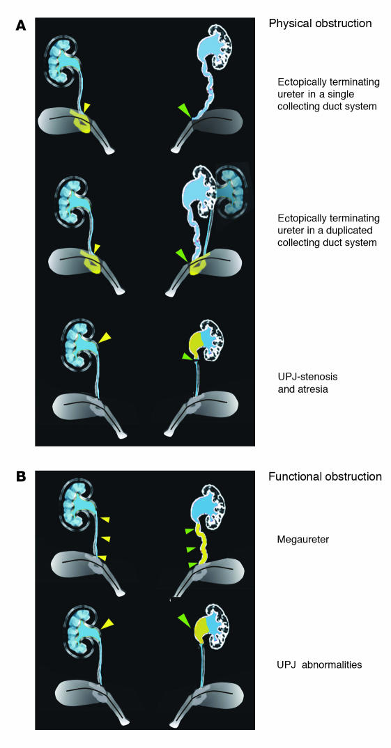
A schematic showing different types of obstruction that can cause hydronephrosis. (A) Top, examples of physical obstruction: ectopically terminating ureter in a single (top) or duplicated (middle) collecting duct system. In both cases the ureter joins the urinary tract outside the normal integration site in the trigone. In the example showing a duplicated system, one ureter joins normally; the other, abnormally. Bottom, uteropelvic junction (UPJ) stenosis or atresia causing physical blockage at the ureteropelvic junction. (B) Examples of functional obstruction. Top, primary megaureter caused by impaired peristalsis or defective differentiation of smooth muscle in the ureter coat. Bottom, UPJ abnormalities caused by failure in outgrowth or function of the renal pelvis. On the left, yellow filled arrowheads designate the normal structure; the abnormal structure on the right is designated by green filled arrowheads.
Comment on
-
Calcineurin is required in urinary tract mesenchyme for the development of the pyeloureteral peristaltic machinery.J Clin Invest. 2004 Apr;113(7):1051-8. doi: 10.1172/JCI20049. J Clin Invest. 2004. PMID: 15057312 Free PMC article.
References
-
- Chevalier RL. Pathophysiology of obstructive nephropathy in the newborn. Semin. Nephrol. 1998;18:585–593. - PubMed
-
- Woolf, A.S., Winyard, P.J.D., Hermanns, M.M., and Welham, J.M. 2003. Maldevelopment of the human kidney and lower urinary tract: an overview. In The kidney: from normal development to congenital disease. P.D. Vize, A.S. Woolf, and J.B.L. Bard, editors. Academic Press Inc. Amsterdam, The Netherlands/ Boston, Massachusetts, USA. 377–393.
-
- Kuwayama F, Miyazaki Y, Ichikawa I. Embryogenesis of the congenital anomalies of the kidney and the urinary tract. Nephrol. Dial. Transplant. 2002;17:45–47. - PubMed
-
- Batourina E, et al. Vitamin A controls epithelial/mesenchymal interactions through Ret expression. Nat. Genet. 2001;27:74–78. - PubMed
