

Expression of Notch-1 and its ligand Jagged-1 in rat liver during liver regeneration
- PMID: 15057910
- PMCID: PMC1769555
- DOI: 10.1002/hep.20156
Expression of Notch-1 and its ligand Jagged-1 in rat liver during liver regeneration
Abstract
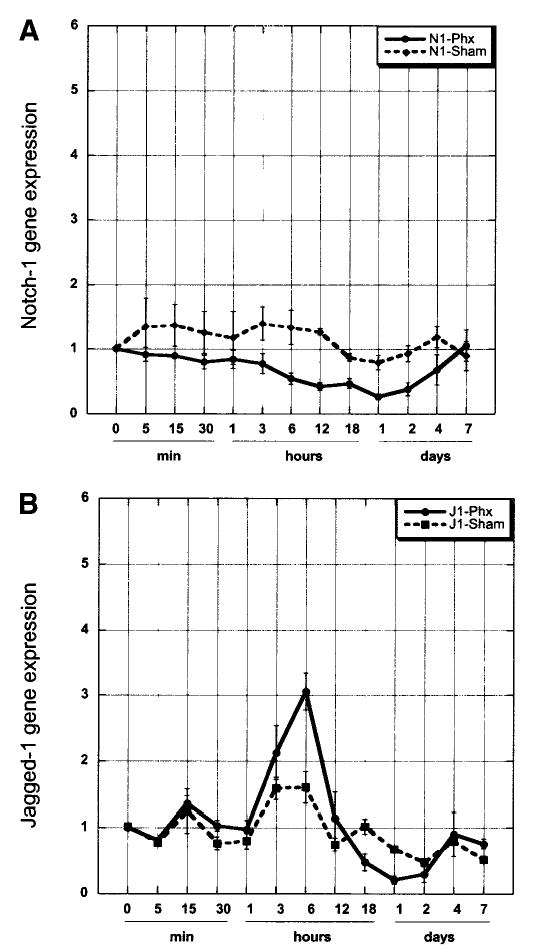
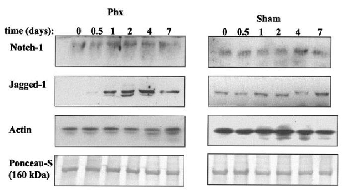
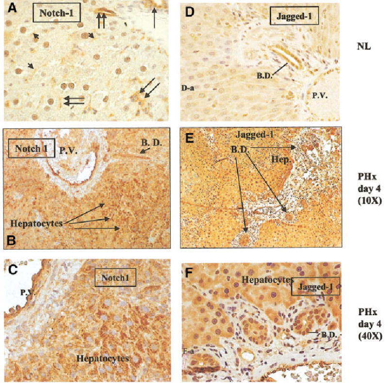
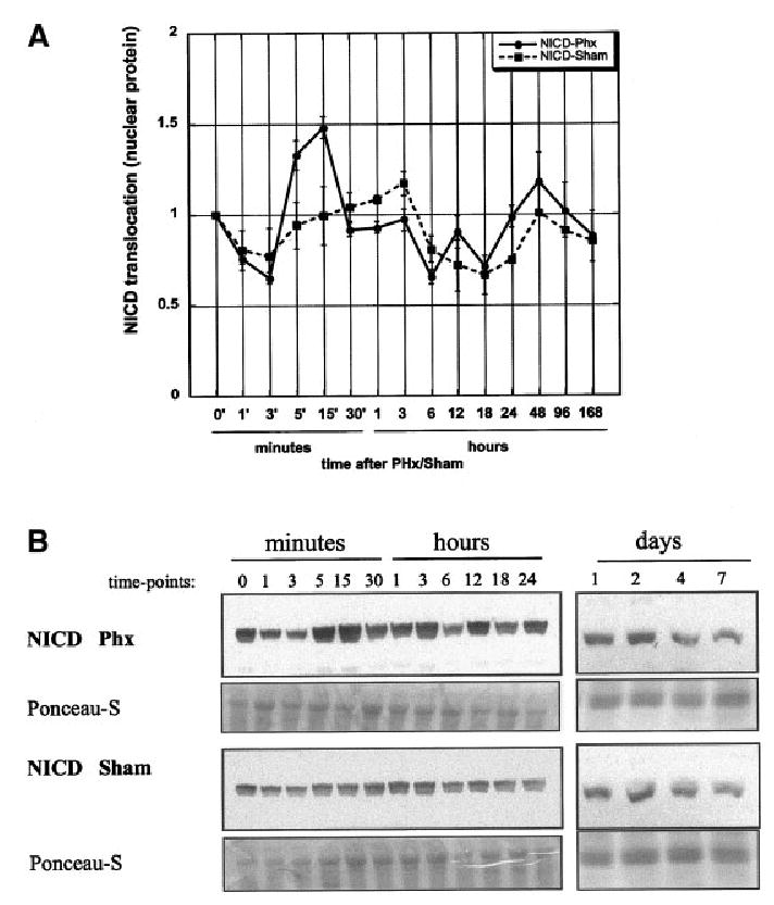
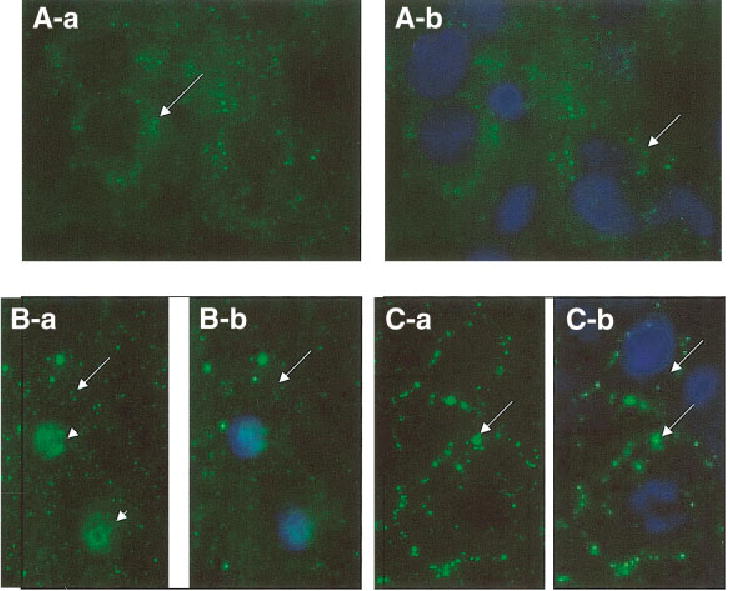
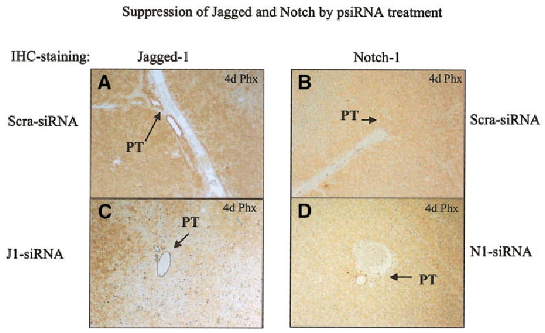
The Notch/Jagged signaling pathway is important for cellular differentiation and proliferation. Its dysfunction is associated with human pathologies in several tissues including liver. Point mutations in Jagged-1 gene are the cause for Alagille syndrome, associated with paucity of intrahepatic bile ducts. To determine the putative role of the trans-membrane receptor Notch and its ligand Jagged-1 in liver regeneration, we investigated the expression of Notch and Jagged-1 in rat liver following 2/3 partial hepatectomy. Immunohistochemical staining of normal rat liver showed that Notch was expressed in hepatocytes, bile duct cells and endothelial cells, whereas Jagged-1 was expressed in bile duct cells and hepatocytes. Both Notch-1 and Jagged-1 proteins were upregulated in hepatocytes after partial hepatectomy up to day 4. After partial hepatectomy, nuclear translocation of the intracellular cytoplasmic domain of Notch (NICD) increased and peaked within 15 minutes, indicating the activation of Notch. Expression of the Notch-dependent target gene (HES-1) expression increased within 30-60 minutes. Addition of recombinant Jagged-1 protein to primary cultures of hepatocytes stimulated hepatocyte DNA synthesis. Furthermore, injection of silencing RNA for Notch and Jagged-1 to livers 2 days before partial hepatectomy significantly suppressed proliferation of hepatocytes at days 2 to 4 of the regenerative response. In conclusion, Notch/Jagged signaling pathway is activated during liver regeneration and is potentially contributing to signals affecting cell growth and differentiation.
Figures
Similar articles
-
The Jagged-1/Notch-1/Hes-1 pathway is involved in intestinal adaptation in a massive small bowel resection rat model.Dig Dis Sci. 2013 Sep;58(9):2478-86. doi: 10.1007/s10620-013-2680-3. Epub 2013 Apr 18. Dig Dis Sci. 2013. PMID: 23595520
-
Alterations of Notch/Jagged mRNA and protein expression after partial hepatectomy in rats.Scand J Gastroenterol. 2008;43(12):1522-8. doi: 10.1080/00365520802321188. Scand J Gastroenterol. 2008. PMID: 18781453
-
Notch signaling controls hepatoblast differentiation by altering the expression of liver-enriched transcription factors.J Cell Sci. 2004 Jul 1;117(Pt 15):3165-74. doi: 10.1242/jcs.01169. J Cell Sci. 2004. PMID: 15226394
-
Notch signalling beyond liver development: emerging concepts in liver repair and oncogenesis.Clin Res Hepatol Gastroenterol. 2013 Nov;37(5):447-54. doi: 10.1016/j.clinre.2013.05.008. Epub 2013 Jun 24. Clin Res Hepatol Gastroenterol. 2013. PMID: 23806629 Review.
-
Notch signaling in vascular development.Arterioscler Thromb Vasc Biol. 2003 Apr 1;23(4):543-53. doi: 10.1161/01.ATV.0000060892.81529.8F. Epub 2003 Feb 13. Arterioscler Thromb Vasc Biol. 2003. PMID: 12615665 Review.
Cited by
-
The Jagged-1/Notch-1/Hes-1 pathway is involved in intestinal adaptation in a massive small bowel resection rat model.Dig Dis Sci. 2013 Sep;58(9):2478-86. doi: 10.1007/s10620-013-2680-3. Epub 2013 Apr 18. Dig Dis Sci. 2013. PMID: 23595520
-
Liver regeneration, growth factors, and amphiregulin.Gastroenterology. 2005 Feb;128(2):503-6. doi: 10.1053/j.gastro.2004.12.039. Gastroenterology. 2005. PMID: 15685562 Free PMC article. No abstract available.
-
Sox2 and JAGGED1 expression in normal and drug-damaged adult mouse inner ear.J Assoc Res Otolaryngol. 2008 Mar;9(1):65-89. doi: 10.1007/s10162-007-0106-7. Epub 2007 Dec 22. J Assoc Res Otolaryngol. 2008. PMID: 18157569 Free PMC article.
-
Exosomes from liver progenitor cells carrying JAG1 activate notch signaling to promote liver regeneration in PVL rats.Cell Death Dis. 2025 Aug 12;16(1):609. doi: 10.1038/s41419-025-07925-1. Cell Death Dis. 2025. PMID: 40796539 Free PMC article.
-
Oxidative stress-induced Notch1 signaling promotes cardiogenic gene expression in mesenchymal stem cells.Stem Cell Res Ther. 2013 Apr 18;4(2):43. doi: 10.1186/scrt190. Stem Cell Res Ther. 2013. PMID: 23597145 Free PMC article.
References
-
- Grisham J. A morphologic study of deoxyribonucleic acid synthesis and cell proliferation in regenerating liver; autoradiography with thymidine-H3. Cancer Res. 1962;22:842–849. - PubMed
-
- Michalopoulos GK, DeFrances MC. Liver regeneration. Science. 1997;276:60 –66. - PubMed
-
- Baron M, Aslam H, Flasza M, Fostier M, Higgs JE, Mazaleyrat SL, Wilkin MB. Multiple levels of Notch signal regulation (review) Mol Membr Biol. 2002;19:27–38. - PubMed
-
- Baron M. An overview of the Notch signalling pathway. Semin Cell Dev Biol. 2003;14:113–119. - PubMed
-
- Artavanis-Tsakonas S, Rand MD, Lake RJ. Notch signaling: cell fate control and signal integration in development. Science. 1999;284:770 –776. - PubMed
Publication types
MeSH terms
Substances
Grants and funding
LinkOut - more resources
Full Text Sources
Molecular Biology Databases