Review
. 2004 Apr;26(4):374-86.
doi: 10.1002/bies.20009.

Anterior eye development and ocular mesenchyme: new insights from mouse models and human diseases

Affiliations
Review

Anterior eye development and ocular mesenchyme: new insights from mouse models and human diseases

Ales Cvekl et al. Bioessays. 2004 Apr.

Abstract

During development of the anterior eye segment, cells that originate from the surface epithelium or the neuroepithelium need to interact with mesenchymal cells, which predominantly originate from the neural crest. Failures of proper interaction result in a complex of developmental disorders such Peters' anomaly, Axenfeld-Rieger's syndrome or aniridia. Here we review the role of transcription factors that have been identified to be involved in the coordination of anterior eye development. Among these factors is PAX6, which is active in both epithelial and mesenchymal cells during ocular development, albeit at different doses and times. We propose that PAX6 is a key element that synchronizes the complex interaction of cell types of different origin, which are all needed for proper morphogenesis of the anterior eye. We discuss several molecular mechanisms that might explain the effects of haploinsufficiency of PAX6 and other transcription factors, and the broad variation of the resulting phenotypes.


Figures

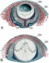
Figure 1
Figure 1
Schematic diagram of ocular mesenchyme development in the mouse eye between embryonic days (E) 12.5-14.5. A: At E 12.5-13.5, the lens vesicle (LV) has detached from the surface epithelium (SE) and has become invaginated into the optic cup. Mesenchymal cells (ME) start to migrate into the space between the anterior epithelium of the lens vesicle and the surface ectoderm. The inner layer of the optic cup forms the neural retina (Re), the outer layer the retinal pigmented epithelium (PE). The optic cup is incompletely inferior at the so-called embryonic (choroidal) fissure (EF), where the hyaloid artery (HA) enters the optic cup. B: At E 13.5-14.5, the mesenchyme cells condense to form several flat layers that are separated from each other by a loose fibrillar extracellular matrix. In the lens (Le), the primary lens fibers elongate to close the lumen of the lens vesicle.
Figure 2
Figure 2
Schematic diagram of ocular mesenchyme development in the mouse eye between embryonic days (E) 14.5 and 19.5. A: At E14.5-15.5, the posterior mesenchyme cells closest to the lens flatten, become connected by apicolateral contacts and form an endothelial monolayer. At the end of this process, all layers of the future cornea have been defined. The endothelial monolayer that has been formed from posterior mesenchyme cells will become the corneal endothelium (CEn), the surface ectoderm that covers the anterior side of the mesenchyme will become the corneal epithelium (CEp). Mesenchyme cells between the corneal epithelium and endothelium differentiate into keratocytes, the specific cell type of the corneal stroma. During differentiation of the corneal endothelium, the lens (L) detaches from the future cornea and a fluid-filled cavity, the anterior chamber (AC), is generated between both structures. In parallel, a new group of mesenchyme cells (Me) arrives at the angle between the future cornea and the anterior edge of the optic cup. B: Beginning at approx. E 15.5, the anterior edge of the optic cup enlarges to form the iris and ciliary body. Mesenchyme cells migrate along the epithelial layers of both structures and finally differentiate into the stroma of the iris (SIr) and ciliary body (SCB). Re: Medina, PE: pigmented epithelium.
Figure 3
Figure 3
Schematic diagram of the development of chamber angle and trabecular meshwork in the mouse eye between postnatal days (P) 1 and 14. A: From P1-P4, the chamber angle is occupied by a dense mass of mesenchymal cells (arrows). B: From P4-P10, chamber angle cells (solid arrows) become separated from each other by small open spaces that are partially filled with extracellular fibers, while vessels appear in the immediate adjacent sclera (open arrows). During this period, the chamber angle is level with the anterior border of the future trabecular meshwork. C: From P11-P14, the extracellular fibers in the chamber angle organize themselves into trabecular beams that become covered by trabecular meshwork cells, while the scleral vessels next to the chamber angle coalesce to Schlemm’s canal. In parallel, the peripheral margin of the anterior chamber moves posteriorly and the inner surface of the trabecular meshwork becomes exposed to the anterior chamber (AC). Re: Medina, CB: ciliary body.
Figure 4
Figure 4
Schematic drawing of various transcription factors that are involved during anterior eye development. Important functional domains are boxed in colors. DNA-binding domains: PD, paired domain; PD5a, alternatively spliced paired domain; HDBcd, bicoid type homeodomain; HDPrd, paired type homeodomain; FH, forkhead domain; bZIP, basic leucine zipper domain. Transcriptional activation domains are shown as green boxes.
Figure 5
Figure 5
Different Pax6 roles in ocular cell differentiation. A high and continuous expression of Pax6 in cells of ectodermal origin (lens, corneal epithelium, iris, and ciliary epithelium) is required for expression of transcription factors (Six3, c-Maf and Prox1),(62,63) structural genes (crystallins and cell adhesion molecules),(65-69) and signaling molecules affecting the migration of neural crest cells into the eye by inductive processes.(61) In addition, a low and transient expression of Pax6 plays cell autonomous roles in the differentiation of trabecular meshwork, and in the formation of corneal endothelium and keratocytes.(53)
Figure 6
Figure 6
Molecular models explaining Pax6 haploinsufficiency and overexpression. A: Interaction of Pax6 and mutated Pax6 (mut-Pax6) with target genes. A “high-affinity”Pax6-binding site in gene A is occupied in Pax6+/+ and Pax6+/- cells. A “low-affinity” Pax6-binding site in gene B is only occupied in Pax6+/+ cells. If a mutated Pax6 allele encodes for a nonfunctional protein or no protein at all, the low-affinity site(s) remain(s) free. B: Truncated proteins capable of binding to DNA can compete with the normal Pax6 protein as a classical dominant-negative mutation.(83) C: Some mutated Pax6 proteins may sequester a hypothetical auxiliary factor (P6X) from Pax6, thereby blocking its function. D: Overexpression of Pax6 (+/+/n, n=1-5) generates enough of Pax6 protein to saturate low-affinity binding sites, but may disrupt the equilibrium between Pax6 and the hypothetical P6X. P6X in panels C and D may be the same or different hypothetical protein. Proteins modulating Pax6 activity include pRb, c-Maf, Six3, Sox2, Mitf, and others.

References

    1. Duke-Elder S, Cook C. In: Normal and Abnormal Development. Part 1. Embryology. Duke-Elder S, editor. Henry Kimpton; London: 1963.
    1. Haustein J. On the ultrastructure of the developing and adult mouse corneal stroma. Anat Embryol (Berl) 1983;168:291–305. - PubMed
    1. Pei YF, Rhodin JAG. The prenatal development of the mouse eye. Anat Rec. 1970;168:105–126. - PubMed
    1. Düblin I. Comparative embryologic studies of the early development of the cornea and the pupillary membrane in reptiles, birds and mammals. Acta Anat (Basel) 1970;76:381–408. - PubMed
    1. Kidson SH, Kume T, Deng K, Winfrey V, Hogan BL. The forkhead/winged-helix gene, Mf1, is necessary for the normal development of the cornea and formation of the anterior chamber in the mouse eye. Dev Biol. 1999;211:306–322. - PubMed

Publication types

MeSH terms

LinkOut - more resources