Review
. 2004;5(4):219.
doi: 10.1186/gb-2004-5-4-219. Epub 2004 Mar 31.

The annexins

Affiliations
Review

The annexins

Stephen E Moss et al. Genome Biol. 2004.

Abstract

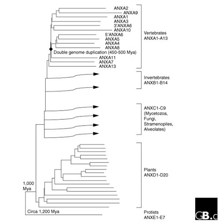
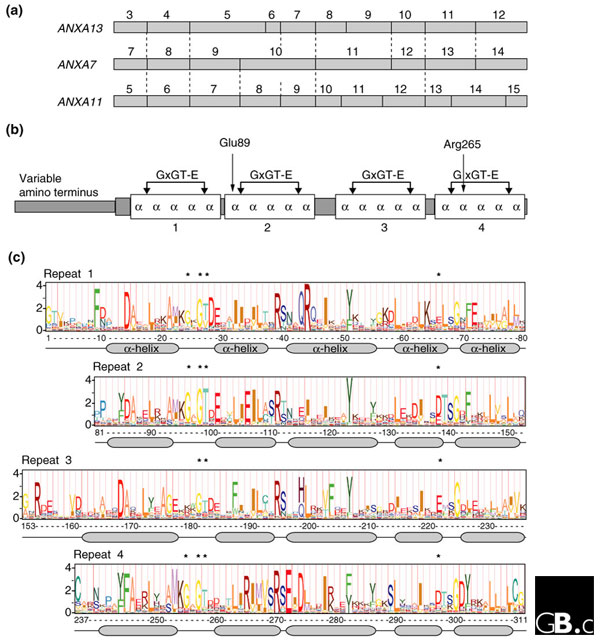
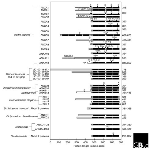
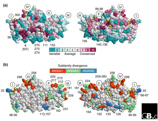
Annexins are traditionally thought of as calcium-dependent phospholipid-binding proteins, but recent work suggests a more complex set of functions. More than a thousand proteins of the annexin superfamily have been identified in major eukaryotic phyla, but annexins are absent from yeasts and prokaryotes. The unique annexin core domain is made up of four similar repeats approximately 70 amino acids long, each of which usually contains a characteristic 'type 2' motif for binding calcium ions. Animal and fungal annexins also have non-homologous amino-terminal domains of varying length and sequence, which are responsible for the distinct localizations and specialized functions of the proteins through post-translational modification and binding to other proteins. Annexins interact with various cell-membrane components that are involved in the structural organization of the cell, intracellular signaling by enzyme modulation and ion fluxes, growth control, and they can act as atypical calcium channels. Analysis of site-specific conservation in the core domain suggests a role for certain buried residues in the calcium-channel activity of vertebrate annexins and in the structural stability of their core domains. Evolutionarily significant differences between subfamilies are preferentially localized to accessible sites on the protein surface that determine membrane binding and interactions with cytosolic proteins.


Figures

Figure 1
Figure 1
The phylogenetic distribution of annexins. A tree showing the classification of annexins into five families, ANXA to ANXE, which correspond with different eukaryotic lineages that originated at different periods over the past 1,200 million years (Mya, million years ago). Names of the vertebrate annexins are shown, but those of other members of the superfamily are omitted for simplicity.
Figure 2
Figure 2
Gene structures, protein domains and signature logos of vertebrate annexins. (a) The organization of the regions of family-A annexin genes encoding the core carboxy-terminal region. Exon numbers are shown above each gene; introns are indicated by vertical lines and homologous intron positions by dotted lines. The structures of the nine human annexin genes not shown are the same as that of ANXA11. ANXA13 is thought to have the gene structure closest to the ancestral vertebrate annexin gene; ANXA7 is intermediate between ANXA13 and the others, and most closely resembles ANXA11 in its amino-terminal half and ANXA13 in its carboxy-terminal half. (b) Annexin proteins generally consist of a unique amino-terminal region (of 0-191 amino acids in vertebrates, for example) and a carboxy-terminal 'core region' of four homologous repeats, each 68-69 amino acids long and containing five α helices and a type-2 calcium binding site with the sequence GxGT-[38 residues]-D/E. The indicated residues Glu89 and Arg265 are considered key components of the putative calcium channel function. (c) Sequence logo for the core domain of vertebrate annexins, derived from a hidden Markov model [11] generated from an alignment of 311 amino acids from 200 sequences representing the 12 subfamilies in 50 vertebrate species. The full height of each residue stack reflects the conservation level at that position; the height of symbols within the stack indicates the relative frequency of each amino acid [12]. The two parts of the calcium-binding motif (GxGT and D/E) are indicated by asterisks. The four repeats are aligned to the right on their calcium-binding motifs.
Figure 3
Figure 3
Domain structures of representative annexin proteins. Orthologs of the 12 human annexins shown in other vertebrates have the same structures, with strict conservation of the four repeats in the core region (black) and variation in length and sequence in the amino-terminal regions (shaded). Human ANXA1 and ANXA2 are shown as dimers, with the member of the S100 protein family that they interact with. Domain structures for other model organisms are derived from public data made available by the relevant genome-sequencing projects. Features: S100Ax, sites for attachment of the indicated member of the S100 family of calcium-binding proteins; P, known phosphorylation sites; K, KGD synapomorphy (a conserved, inherited characteristic of proteins); I, codon insertions (+x denotes the number of codons inserted); S-A/b, nonsynonymous coding polymorphisms (SNPs) with the amino acid in the major variant (A) and that in the minor variant (b); N, putative nucleotide-binding sites; D, codon deletions (-x denotes the number of codons deleted); A, alternatively spliced exons; Myr, myristoylation. The total length of each protein is indicated on the right.
Figure 4
Figure 4
Surface mapping of important sites onto the three-dimensional structure of annexins. All panels show the crystal structure of the core region of the pig annexin A1 protein (Protein Data Bank code:1MCX [19]), viewed frontally (left) and laterally inverted (right) as a space-filling model rendered by the RasTop 2.0 version of RasMol [24]. Residues are numbered as in Figure 2, and the approximate positions of the conserved repeats are indicated with Roman numerals. (a) Functionally important sites common to all annexins. The level of evolutionary conservation in clusters of residues is indicated by lighter or darker shading. This is derived from a maximum-likelihood analysis of a multiple sequence alignment from 200 vertebrate A-family annexins using CONSURF [22,23] NT, amino terminus. (b) Sites that are functionally divergent between annexin subfamilies are shown with different shading for ANXA1, ANXA2 or ANXA5, three annexins for which the differences are especially significant. The sites were assessed by 'rate-shift analysis' of subfamily sequence alignments using DIVERGE [21] RATE4SITE and CONSURF [22,23]. Calcium atoms are indicated by Ca.

References

    1. Creutz CE, Pazoles CJ, Pollard HB. Identification and purification of an adrenal medullary protein (synexin) that causes calcium-dependent aggregation of isolated chromaffin granules. J Biol Chem. 1978;253:2858–2866. The first isolation of a protein of the annexin family (now called annexin A7) and demonstration of its role in vesicle fusion. - PubMed
    1. Huang KS, Wallner BP, Mattaliano RJ, Tizard R, Burne C, Frey A, Hession C, McGray P, Sinclair LK, Chow EP, et al. Two human 35 kD inhibitors of phospholipase A2 are related to substrates of pp60v-src and of the epidermal growth factor receptor/kinase. Cell. 1986;46:191–199. Two proteins, now known as annexins A1 and A2, that were originally characterized as inhibitors of phospholipase A2. - PubMed
    1. Saris CJ, Tack BF, Kristensen T, Glenney JR, Jr, Hunter T. The cDNA sequence for the protein-tyrosine kinase substrate p36 (calpactin I heavy chain) reveals a multidomain protein with internal repeats. Cell. 1986;46:201–212. One of the first annexins to be cloned; it derived its name from its ability to bind calcium and bundle F-actin. - PubMed
    1. Human Gene Nomenclature Committee http://www.gene.ucl.ac.uk/nomenclature/ The official nomenclature source of the Human Gene Organization (HUGO) for human genes and their cognate orthologs.
    1. Gerke V, Moss SE. Annexins: from structure to function. Physiol Rev. 2002;82:331–371. A comprehensive review on annexins. - PubMed

Publication types

LinkOut - more resources