Multicenter Study
. 2004;6(2):R142-50.
doi: 10.1186/ar1149. Epub 2004 Feb 5.

Rheumatoid arthritis specific anti-Sa antibodies target citrullinated vimentin

Affiliations
Multicenter Study

Rheumatoid arthritis specific anti-Sa antibodies target citrullinated vimentin

Erik R Vossenaar et al. Arthritis Res Ther. 2004.

Abstract

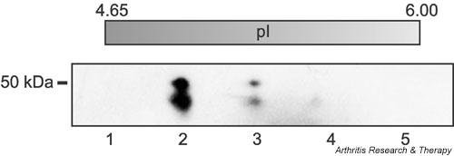
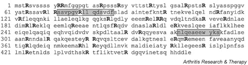
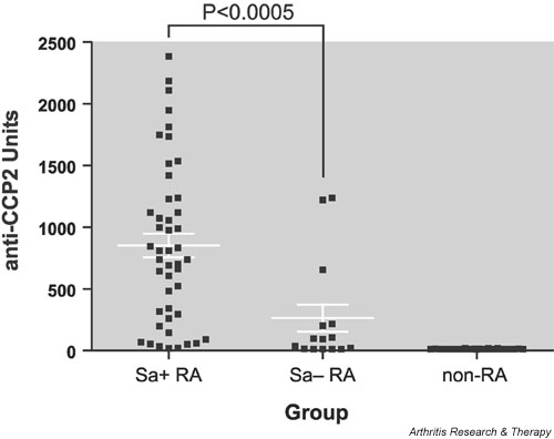
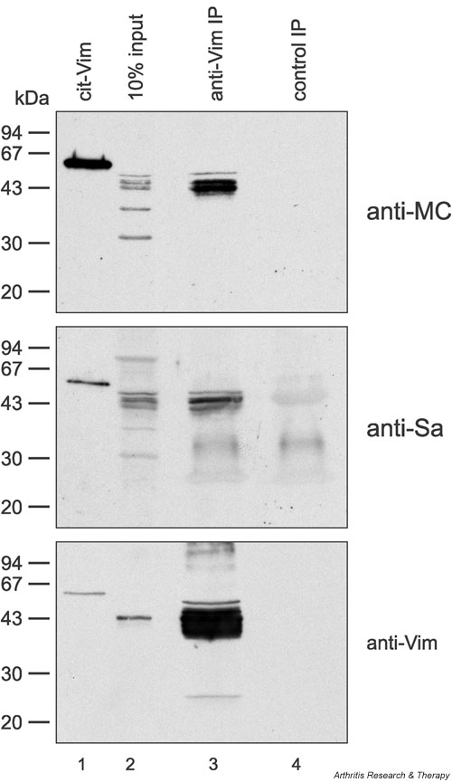
Antibodies directed to the Sa antigen are highly specific for rheumatoid arthritis (RA) and can be detected in approximately 40% of RA sera. The antigen, a doublet of protein bands of about 50 kDa, is present in placenta and in RA synovial tissue. Although it has been stated that the Sa antigen is citrullinated vimentin, experimental proof for this claim has never been published. In this study, we investigated the precise nature of the antigen. Peptide sequences that were obtained from highly purified Sa antigen were unique to vimentin. Recombinant vimentin, however, was not recognized by anti-Sa reference sera. In vivo, vimentin is subjected to various post-translational modifications, including citrullination. Since antibodies to citrullinated proteins are known to be highly specific for RA, we investigated whether Sa is citrullinated and found that Sa indeed is citrullinated vimentin. Anti-Sa antibodies thus belong to the family of anticitrullinated protein/peptide antibodies. The presence of the Sa antigen in RA synovial tissue, and the recent observation that vimentin is citrullinated in dying human macrophages, make citrullinated vimentin an interesting candidate autoantigen in RA and may provide new insights into the potential role of citrullinated synovial antigens and the antibodies directed to them in the pathophysiology of RA.


Figures

Figure 1
Figure 1
Purification of placental Sa antigen. The antigen was purified by anion-exchange chromatography from extracts of placenta and subsequently purified by two-dimensional gel electrophoresis according to a three-step procedure described by Liang and colleagues [20]. First, proteins were separated by molecular weight, then proteins of appropriate molecular weight were separated by isoelectric focusing (IEF), and finally proteins with appropriate pI were separated once more by molecular weight. Each step of the procedure was monitored by western blotting with an anti-Sa reference serum. Shown here is the final gel, which was stained with Coomassie brilliant blue. The double band in lane 2 is the Sa antigen that was cut out and used for microsequencing. Each lane represents a portion of the IEF gel (approximate pI is listed above each lane).
Figure 2
Figure 2
Amino acid sequence of vimentin with Sa microsequences and sequence of human vimentin (Swiss-Prot database number P08670). Two distinct peptides that were obtained by microsequencing are indicated in grey. Peptide 72–86 was obtained twice, VD84–85 was ambiguous in one of the peptides, and R78 could not be sequenced in both peptides. All arginines are given in capital and bold, because they can potentially be modified to citrulline by peptidylarginine deiminase.
Figure 3
Figure 3
Comparison of anti-CCP titers in Sa+ and Sa- patients. To investigate a possible relationship between anti-Sa and anti-CCP autoantibodies, we compared anti-CCP2 antibody titers in 46 anti-Sa-positive rheumatoid arthritis (RA) sera, 15 anti-Sa-negative RA sera, and 26 control sera, using the CCP2 test kit. Ninety-six percent of anti-Sa-positive RA patients, 60% of Sa-negative RA-patients, and none of the control patients was positive for anti-CCP2. Anti-Sa-positive RA sera had a significantly higher anti-CCP titer (852 ± 96 U; mean ± SEM) than anti-SA-negative sera (263 ± 110 U) (P < 0.0005; Mann–Whitney test). None of the control sera tested positive in either of the two assays (12 ± 0.4 U).
Figure 4
Figure 4
Placental Sa antigen is citrullinated vimentin. Three identical immunoblots containing semipurified placental Sa antigen (100 μg; lanes 1, 4, 7), human recombinant vimentin (Vim) (50 ng; lanes 2, 5, 8), and human recombinant vimentin that had been citrullinated in vitro (cit-Vim) (50 ng; lanes 3, 6, 9) were stained with either anti-Sa reference serum (left panel), anti-modified citrulline (anti-MC) antibodies (middle panel), or antivimentin (anti-Vim) antibodies (right panel). Sa antigen is recognized both by anti-MC antibodies and by anti-Vim antibodies (lanes 4 and 7, respectively), indicating that the antigen is indeed citrullinated vimentin. Citrullinated vimentin was recognized by the anti-Sa serum, whereas unmodified vimentin was not (lanes 3 and 2, respectively), indicating that the presence of citrulline residues is essential for the autoantigenicity of Sa. Molecular weight markers are indicated on the left.
Figure 5
Figure 5
Antivimentin (anti-Vim) can immunoprecipitate Sa antigen from placental extract. Vimentin was immunoprecipitated from semipurified placental extract with monoclonal antibody RV202 (lanes 3). Immunoprecipitated vimentin was stained by anti-modified citrulline (anti-MC) antibodies (upper panel), anti-Sa serum (middle panel), or polyclonal antivimentin (anti-Vim) antibodies (lower panel), indicating that Sa is citrullinated vimentin. An immunoprecipitation (IP) with an isotype-matched monoclonal control antibody 4G3 (lanes 4) served as a negative control. Lanes 1 contain human recombinant vimentin citrullinated in vitro (cit-Vim) (50 ng) as a positive control. Lanes 2 show 10% input. Molecular weight markers are indicated on the left.

Comment in

References

    1. van Boekel MA, Vossenaar ER, van den Hoogen FH, van Venrooij WJ. Autoantibody systems in rheumatoid arthritis: specificity, sensitivity and diagnostic value. Arthritis Res. 2002;4:87–93. doi: 10.1186/ar395. - DOI - PMC - PubMed
    1. Steiner G, Smolen J. Autoantibodies in rheumatoid arthritis and their clinical significance. Arthritis Res. 2002;Suppl 2:S1–S5. doi: 10.1186/ar551. - DOI - PMC - PubMed
    1. Mageed RA. The RF antigen. In: van Venrooij WJ, Maini RN, editor. In Manual of Biological Markers of Disease. Section B1.1. Dordrecht, The Netherlands: Kluwer Academic Publishers; 1996. pp. 1–27.
    1. Lisse JR. Does rheumatoid factor always mean arthritis? Postgrad Med. 1993;94:133–134. - PubMed
    1. Palosuo T, Tilvis R, Strandberg T, Aho K. Filaggrin related antibodies among the aged. Ann Rheum Dis. 2003;62:261–263. doi: 10.1136/ard.62.3.261. - DOI - PMC - PubMed

Publication types