Comparative Study
. 2004 Apr;186(8):2295-302.
doi: 10.1128/JB.186.8.2295-2302.2004.

GldI is a lipoprotein that is required for Flavobacterium johnsoniae gliding motility and chitin utilization

Affiliations
Comparative Study

GldI is a lipoprotein that is required for Flavobacterium johnsoniae gliding motility and chitin utilization

Mark J McBride et al. J Bacteriol. 2004 Apr.

Abstract

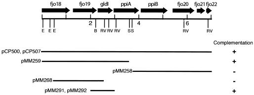
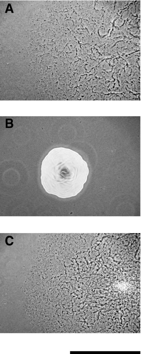
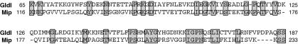
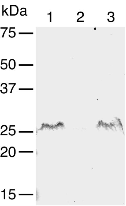
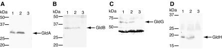
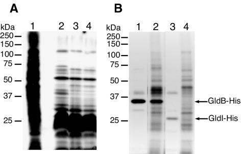
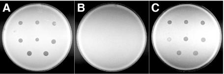
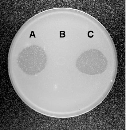
Cells of Flavobacterium johnsoniae glide rapidly over surfaces by an unknown mechanism. Seven genes (gldA, gldB, gldD, gldF, gldG, gldH, and ftsX) that are required for gliding motility have been described. Complementation of the nonmotile mutants UW102-41, UW102-85, and UW102-92 identified another gene, gldI, that is required for gliding motility. gldI mutants formed nonspreading colonies, and individual cells were completely nonmotile. They were also resistant to bacteriophages that infect wild-type cells, and they failed to digest chitin. Introduction of wild-type gldI on a plasmid restored colony spreading, cell motility, phage sensitivity, and the ability to digest chitin to the gldI mutants. gldI encodes a predicted 199-amino-acid protein that localized to the membrane fraction. Labeling studies with [(3)H]palmitate indicated that GldI is a lipoprotein. GldI is similar to peptidyl-prolyl cis/trans-isomerases of the FK506-binding protein family and may be involved in folding cell envelope protein components of the motility machinery.


Figures

FIG. 1.
FIG. 1.
Map of the gldI region of F. johnsoniae. Restriction sites are indicated as follows: B, BglII; E, EcoRI; RV, EcoRV; and S, SacI. Kilobase pair positions 2, 4, and 6 are indicated. The presence (+) or absence (−) of complementation of gldI mutants by fragments cloned into shuttle vectors is indicated beneath the map.
FIG. 2.
FIG. 2.
Photomicrographs of F. johnsoniae colonies. Colonies were grown for 30 h at 25°C on PY2 agar medium. Photomicrographs were taken with a Kodak DC290 digital camera mounted on an Olympus IMT-2 inverted microscope. (A) Wild-type F. johnsoniae UW101; (B) gldI mutant UW102-41; (C) UW102-41 complemented with pMM291, which carries gldI. Bar, 1 mm.
FIG. 3.
FIG. 3.
Alignment of F. johnsoniae GldI sequence with L. pneumophila Mip sequence. Identical residues are boxed and shaded. The numbers are amino acid residue positions starting from the amino terminus of each protein. Full-length GldI has 218 amino acids, and Mip has 233 amino acids. Gaps introduced to maximize alignment are indicated by dashes.
FIG. 4.
FIG. 4.
Localization of GldI-His. Cells of F. johnsoniae UW102-41 expressing GldI-His from pTB45 were fractionated, and GldI-His was detected by Western blot analysis. Lane 1, whole cells; lane 2, soluble fraction; lane 3, membrane fraction. Equal amounts of protein were loaded in each lane.
FIG. 5.
FIG. 5.
Effect of mutation in gldI on other Gld proteins. Cell extracts (20 μg of protein per lane) were examined for GldA (A), GldB (B), GldG (C), and GldH (D) proteins by Western blot analysis. In panels A to D, lane 1 contains wild-type cells and lane 2 contains cells of the gldI mutant UW102-41, while lane 3 contains cells of the gldA mutant F. johnsoniae CJ288 (A), cells of the gldB mutant F. johnsoniae CJ569 (B), cells of the gldFG mutant F. johnsoniae CJ787 (C), and cells of the gldH mutant F. johnsoniae CJ1043 (D).
FIG. 6.
FIG. 6.
GldI is a lipoprotein. (A) Cells of F. johnsoniae were labeled with either [3H]glutamate (to label nearly all proteins) or [3H]palmitate (to label lipoproteins). Proteins were separated by SDS-PAGE and detected by autoradiography. Lane 1, wild-type cells labeled with [3H]glutamate; lane 2, wild-type cells labeled with [3H]palmitate; lane 3, cells of the gldI mutant UW102-41 labeled with [3H]palmitate; lane 4, cells of UW102-41 complemented with pMM291 (which carries gldI) labeled with [3H]palmitate. (B) Radiolabeling of GldB-His and GldI-His. Cells of F. johnsoniae were labeled with [3H]palmitate or [3H]glutamate. Proteins were isolated by precipitation with Ni-NTA His-Bind resin, separated by SDS-PAGE, and detected by autoradiography. Lane 1, cells expressing GldB-His from pTB39, labeled with [3H]palmitate; lane 2, cells expressing GldB-His from pTB39, labeled with [3H]glutamate; lane 3, cells expressing GldI-His from pTB45, labeled with [3H]palmitate; lane 4, cells expressing GldI-His from pTB45, labeled with [3H]glutamate.
FIG. 7.
FIG. 7.
Effect of mutation in gldI on bacteriophage resistance. Bacteriophages (2.5 μl of lysates containing approximately 6 × 10⁷ phage/ml) were spotted onto lawns of cells in CYE overlay agar. Bacteriophages were spotted onto three strains of F. johnsoniae, the wild-type strain (A), the gldI mutant strain UW102-41 (B), and strain UW102-41 complemented with pMM291 (which carries gldI) (C). The plates were incubated at 25°C for 24 h to observe lysis. Bacteriophages were applied to the bacteria in the following order from left to right: for the top row, φCj1, φCj13, and φCj23; for the middle row, φCj28, φCj29, and φCj42; for the bottom row, φCj48 and φCj54. The diameter of the petri dish is 9 cm.
FIG. 8.
FIG. 8.
Effect of mutation in gldI on the ability to utilize chitin. Approximately 4 × 10⁷ cells of wild-type F. johnsoniae (A), the gldI mutant strain UW102-41 (B), and strain UW102-41 complemented with pMM291 (which carries gldI) (C) were spotted on MYA-chitin medium and incubated for 6 days at 25°C.

References

    1. Agarwal, S., D. W. Hunnicutt, and M. J. McBride. 1997. Cloning and characterization of the Flavobacterium johnsoniae (Cytophaga johnsonae) gliding motility gene, gldA. Proc. Natl. Acad. Sci. USA 94:12139-12144. - PMC - PubMed
    1. Altschul, S. F., W. Gish, W. Miller, E. W. Myers, and D. J. Lipman. 1990. Basic local alignment search tool. J. Mol. Biol. 215:403-410. - PubMed
    1. Buell, C., V. Joardar, M. Lindeberg, J. Selengut, I. Paulsen, M. Gwinn, R. Dodson, R. Deboy, A. Durkin, J. Kolonay, R. Madupu, S. Daugherty, L. Brinkac, M. Beanan, D. Haft, W. Nelson, T. Davidsen, N. Zafar, L. Zhou, J. Liu, Q. Yuan, H. Khouri, N. Fedorova, B. Tran, D. Russell, K. Berry, T. Utterback, S. Van Aken, T. Feldblyum, M. D'Ascenzo, W. Deng, A. Ramos, J. Alfano, S. Cartinhour, A. Chatterjee, T. Delaney, S. Lazarowitz, G. Martin, D. Schneider, X. Tang, C. Bender, O. White, C. Fraser, and A. Collmer. 2003. The complete genome sequence of the Arabidopsis and tomato pathogen Pseudomonas syringae pv. tomato DC3000. Proc. Natl. Acad. Sci. USA 100:10181-10186. - PMC - PubMed
    1. Calamita, G., W. Bishai, G. Preston, W. Guggino, and P. Agre. 1995. Molecular cloning and characterization of AqpZ, a water channel from Escherichia coli. J. Biol. Chem. 270:29063-29066. - PubMed
    1. Chang, L. Y. E., J. L. Pate, and R. J. Betzig. 1984. Isolation and characterization of nonspreading mutants of the gliding bacterium Cytophaga johnsonae. J. Bacteriol. 159:26-35. - PMC - PubMed

Publication types

Associated data

LinkOut - more resources