Comparative Study
. 2004 Apr;186(8):2402-12.
doi: 10.1128/JB.186.8.2402-2412.2004.

Genetic analysis of the Salmonella enterica type III secretion-associated ATPase InvC defines discrete functional domains

Affiliations
Comparative Study

Genetic analysis of the Salmonella enterica type III secretion-associated ATPase InvC defines discrete functional domains

Yukihiro Akeda et al. J Bacteriol. 2004 Apr.

Abstract

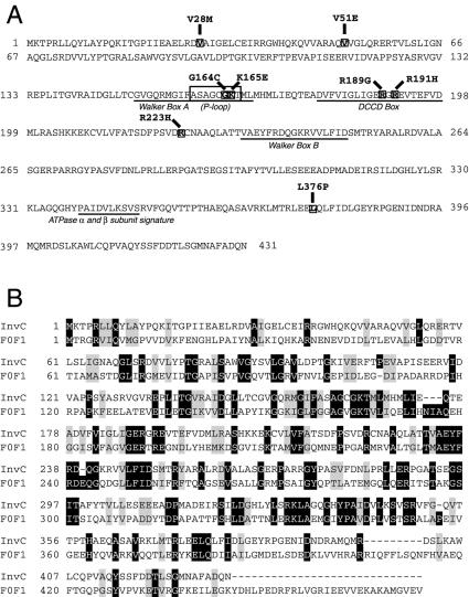
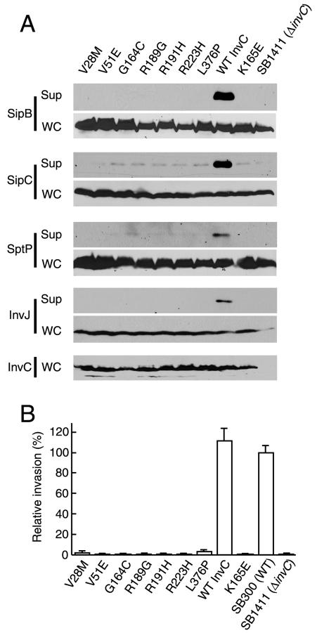
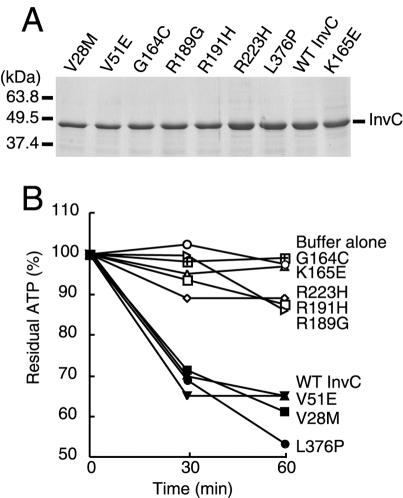
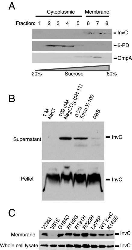
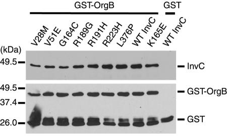
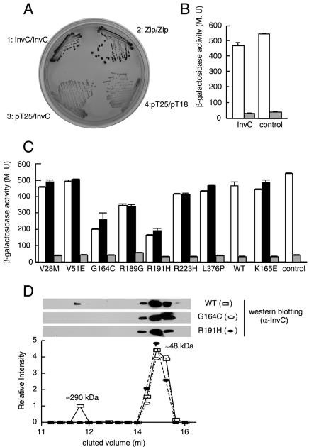
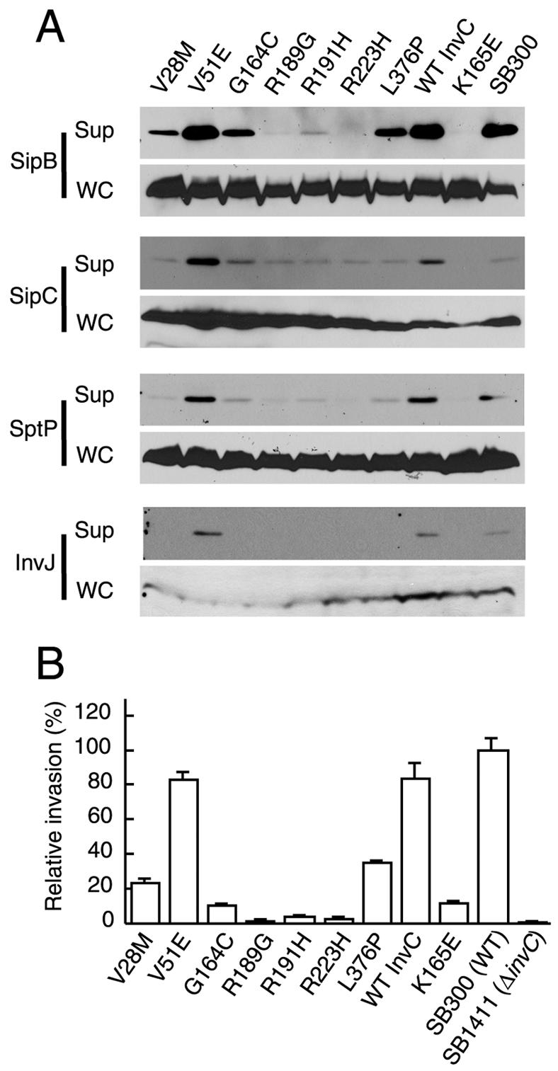
An essential component of all type III secretion systems is a highly conserved ATPase that shares significant amino acid sequence similarity to the beta subunit of the F(0)F(1) ATPases and is thought to provide the energy for the secretion process. We have performed a genetic and functional analysis of InvC, the ATPase associated with the Salmonella enterica type III secretion system encoded within its pathogenicity island 1. Through a mutagenesis analysis, we have identified amino acid residues that are essential for specific activities of InvC, such as nucleotide hydrolysis and membrane binding. This has allowed us to define discrete domains of InvC that are specifically associated with different essential activities of this protein.


Figures

FIG. 1.
FIG. 1.
Locations of loss-of-function mutations in InvC in its primary amino acid sequence. (A) Locations of known predicted motifs within the primary amino acid sequence of InvC. Walker boxes A and B, the dicyclohexylcarbodiimide-binding site (DCCD box), and the F0F1 α and β subunit signature site are also indicated. (B) Amino acid sequence comparison of InvC with the β subunit of the E. coli F0F1 ATPase. Black boxes denote identity, and grey boxes indicate conservative substitutions.
FIG. 2.
FIG. 2.
Loss-of-function InvC mutants are defective in type III secretion and invasion of cultured cells. (A) Bacterial cultured supernatants (Sup) and whole-cell lysates (WC) of S. enterica serovar Typhimurium strains expressing different invC mutants were examined for their levels of InvC (for whole-cell lysates only) and the presence of the type III secreted proteins SipB, SipC, SptP, and InvJ. (B) Abilities of different strains to invade cultured intestinal Henle-407 cells, as measured by a gentamicin resistance assay. Values (means ± standard deviations) represent percentages of the inocula that resisted gentamicin treatment as a consequence of bacterial invasion and have been normalized to the level of entry of wild-type S. enterica serovar Typhimurium, which was considered 100% (the actual value for the wild type was 4.9 ± 0.43).
FIG. 3.
FIG. 3.
ATPase activity of loss-of-function InvC mutants. Wild-type and mutant InvC proteins were purified to homogeneity, applied to SDS-PAGE gels, and stained with Coomassie brilliant blue to ascertain the purity of the preparations (A). The ATPase activities of the different protein preparations were measured with a luciferase-based assay (see Materials and Methods) (B). Equivalent results were obtained for several repetitions of this assay.
FIG. 4.
FIG. 4.
Membrane association of wild-type InvC and its loss-of-function mutants. (A) Cell lysate from wild-type S. enterica serovar Typhimurium was applied to a sucrose gradient. Fractions were collected, and the presence of InvC, the cytoplasmic protein 6-phosphogluconate dehydrogenase (6-PD), or the membrane protein OmpA was examined by Western immunoblotting using specific antibodies. (B) A crude membrane fraction of wild-type S. enterica serovar Typhimurium was subjected to the indicated treatments, and the presence of InvC in either the pellet (membrane) or supernatant (extracted material) of the treated samples was examined by Western immunoblotting. (C) Whole-cell lysates and crude membrane preparations of S. enterica serovar Typhimurium strains expressing the different invC mutants were examined for the presence of InvC by Western immunoblotting.
FIG. 5.
FIG. 5.
Interaction of InvC and its loss-of-function mutants with the TTSS-associated protein OrgB. The interaction of InvC and its mutants with OrgB was examined by a GST pull-down assay, as indicated in Materials and Methods. Beads loaded with GST or GST-OrgB (as indicated) were added to whole-cell lysates of S. enterica serovar Typhimurium strains expressing either the wild type or different invC mutants. The presence of InvC bound to the beads was examined by Western immunoblotting. Blots were reprobed with an antibody directed to GST to ascertain equal loading of the beads. Notice that reprobed blots of beads loaded with GST-OrgB showed bands corresponding to GST-OrgB as well as breakdown products.
FIG. 6.
FIG. 6.
Homotypic interaction of wild-type InvC and mutant proteins. (A) Wild-type InvC was examined for the ability to interact with itself in a bacterial adenylate cyclase reconstitution two-hybrid assay as a surrogate for its ability to form oligomers. E. coli strains carrying bait and target plasmids encoding InvC or the leucine zipper domain of the yeast transcription activator GCN4 (Zip) as a positive control or strains carrying empty vectors were plated on MacConkey agar plates to visualize interactions. Colonies of bacteria expressing interacting proteins appear red (dark in this figure). (B) β-Galactosidase activities (a measure of protein-protein interactions) of strains carrying bait and target plasmids encoding InvC or the leucine zipper domain of GCN4 (white bars). Controls for this experiment consisted of the bait vector encoding either InvC or the leucine zipper domain of GCN4 cotransformed with an empty target vector (gray bars). MU, Miller units. (C) The interaction of the different InvC loss-of-function mutants with themselves (white bars) or with wild-type InvC (black bars) was also measured by the bacterial two-hybrid assay by measuring the β-galactosidase activities of the indicated strains. The gray bars show the β-galactosidase activities of strains carrying the indicated mutants in the bait plasmid and an empty target vector control. (D) Gel filtration profile of wild-type InvC and the oligomerization-defective mutants InvCG164C and InvCR191H. Purified proteins were loaded on a gel filtration column as indicated in Materials and Methods, and the different fractions were probed for the presence of InvC by Western immunoblotting.
FIG. 7.
FIG. 7.
Dominant-negative effect of invC loss-of-function mutants. Wild-type S. enterica serovar Typhimurium was transformed with plasmids expressing different loss-of-function invC mutants. The resulting strains were tested for the ability to secrete the SPI-1 TTSS proteins SipB, SipC, SptP, and InvJ (A) and for the ability to invade cultured Henle-407 cells (B). Values in panel B represent means ± standard deviations of the percentages of the original inocula that resisted gentamicin treatment as a consequence of bacterial invasion, and they have been normalized to the level of entry of wild-type S. enterica serovar Typhimurium, which was considered 100% (the actual value for the wild type was 5 ± 0.4).
FIG. 8.
FIG. 8.
Structure-based model of InvC and locations of its loss-of-function mutants. The predicted structure of InvC was modeled by SWISS-MODEL, an automated comparative protein modeling server (http://www.expasy.org/swissmod/SWISS-MODEL.html) (35, 36), using the crystal structure of the highly related β subunit of the F0F1 ATPase as a template. The locations of the loss-of-function mutants as well as the hypothetical locations of the functional domains of InvC (indicated as I, II, and III) are shown. The V28M mutation is not shown since this region of InvC was not present in the modeled structure due to primary amino acid sequence divergence between InvC and the β subunit of the F0F1 ATPase. See the text for details.

References

    1. Auvray, F., A. J. Ozin, L. Claret, and C. Hughes. 2002. Intrinsic membrane targeting of the flagellar export ATPase FliI: interaction with acidic phospholipids and FliH. J. Mol. Biol. 318:941-950. - PMC - PubMed
    1. Boyer, P. 1997. The ATP synthase—a splendid molecular machine. Annu. Rev. Biochem. 66:717-749. - PubMed
    1. Brusilow, W. S. A. 1993. Assembly of the Escherichia coli F1F0 ATPase, a large multimeric membrane-bound enzyme. Mol. Microbiol. 9:419-424. - PubMed
    1. Claret, L., S. R. Calder, M. Higgins, and C. Hughes. 2003. Oligomerization and activation of the FliI ATPase central to bacterial flagellum assembly. Mol. Microbiol. 48:1349-1355. - PMC - PubMed
    1. Collazo, C. M., M. K. Zierler, and J. E. Galán. 1995. Functional analysis of the Salmonella typhimurium invasion genes invI and invJ and identification of a target of the protein secretion apparatus encoded in the inv locus. Mol. Microbiol. 15:25-38. - PubMed

Publication types

MeSH terms

LinkOut - more resources