. 2004 Apr;24(8):3157-67.
doi: 10.1128/MCB.24.8.3157-3167.2004.

In vivo dynamics of Swi6 in yeast: evidence for a stochastic model of heterochromatin

Affiliations

In vivo dynamics of Swi6 in yeast: evidence for a stochastic model of heterochromatin

Thierry Cheutin et al. Mol Cell Biol. 2004 Apr.

Abstract

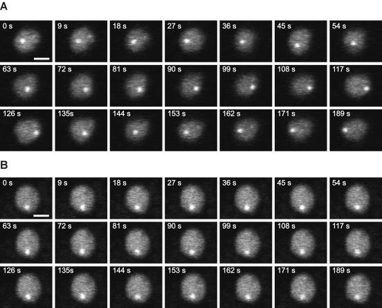
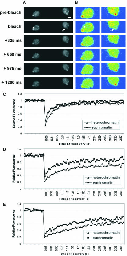
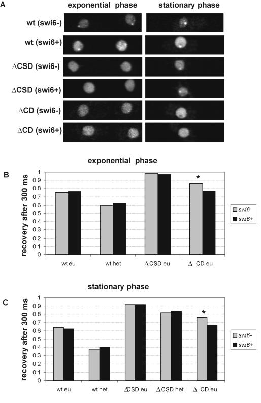
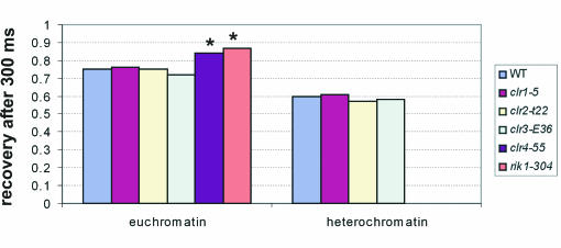
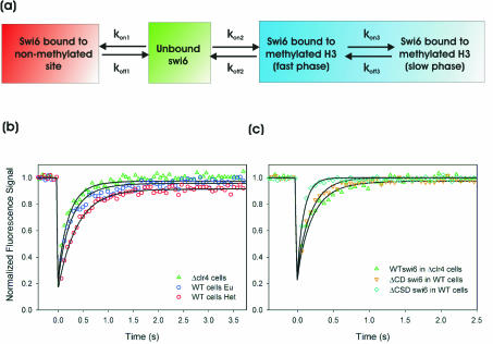
The mechanism for transcriptional silencing of pericentric heterochromatin is conserved from fission yeast to mammals. Silenced genome regions are marked by epigenetic methylation of histone H3, which serves as a binding site for structural heterochromatin proteins. In the fission yeast Schizosaccharomyces pombe, the major structural heterochromatin protein is Swi6. To gain insight into Swi6 function in vivo, we have studied its dynamics in the nucleus of living yeast. We demonstrate that, in contrast to mammalian cells, yeast heterochromatin domains undergo rapid, large-scale motions within the nucleus. Similar to the situation in mammalian cells, Swi6 does not permanently associate with these chromatin domains but binds only transiently to euchromatin and heterochromatin. Swi6 binding dynamics are dependent on growth status and on the silencing factors Clr4 and Rik1, but not Clr1, Clr2, or Clr3. By comparing the kinetics of mutant Swi6 proteins in swi6(-) and swi6(+) strains, we demonstrate that homotypic protein-protein interactions via the chromoshadow domain stabilize Swi6 binding to chromatin in vivo. Kinetic modeling allowed quantitative estimation of residence times and indicated the existence of at least two kinetically distinct populations of Swi6 in heterochromatin. The observed dynamics of Swi6 binding are consistent with a stochastic model of heterochromatin and indicate evolutionary conservation of heterochromatin protein binding properties from mammals to yeast.


Figures

FIG. 1.
FIG. 1.
Time-lapse microscopy of GFP-Swi6. Optical sections were taken from movies collected at the indicated times from yeast growing in exponential phase (A) and stationary phase (B). Heterochromatin domains are highly mobile in the exponential-growth phase, whereas their mobility is greatly reduced during stationary phase. Bar: 1 μm.
FIG. 2.
FIG. 2.
Mobility of GFP-Swi6 in S. pombe. (A) FRAP on exponentially growing yeast cells expressing GFP-Swi6 by bleaching an area in heterochromatin (arrow) or in euchromatin (arrowhead). Bar, 1 μm. (B) Pseudocolor images of those in panel A. (C to E) Comparison of GFP-Swi6 recovery in heterochromatin and euchromatin during exponential phase (C), during stationary phase (D), and after sporulation (E). Values represent averages for 35 cells from five experiments.
FIG. 3.
FIG. 3.
Contribution of the CD and CSD to Swi6 binding. (A) Localization of GFP-Swi6, GFP-ΔCSD, and GFP-ΔCD in swi6 and swi6+ strains growing exponentially or in stationary phase. (B and C) Quantitation of FRAP recovery after 300 ms in swi6 and swi6+ backgrounds for GFP-Swi6-WT, GFP-Swi6ΔCD, and GFP-Swi6ΔCSD during exponential phase (B) and stationary phase (C). Values represent averages for 30 cells from three experiments. *, statistically significant difference in recovery.
FIG. 4.
FIG. 4.
Influence of Clr1, Clr2, Clr3, Clr4, and Rik1 on Swi6 binding. Shown is a quantitation of FRAP recovery after 300 ms in swi6+, clr1-5, clr2-E22, clr3-E36, clr4-S5, and rik1-304 strains during exponential phase. Values represent averages for 30 cells from three experiments. *, statistically significant difference in recovery.
FIG. 5.
FIG. 5.
Kinetic model and corresponding fits to experimental data. (a) Minimal kinetic model for simultaneously fitting Swi6 FRAP data in clr4-S5 cells and in euchromatin and heterochromatin of a WT strain. kon and koff are association and dissociation rate constants, respectively (see Fig. S2 in the supplemental material for a full model). (b) Best fits, determined by least-squares regression analysis, to the model for WT Swi6 recovery in clr4-S5 cells and WT cells in euchromatin and heterochromatin. All parameters are listed in Table 1. Note that all rate constants are the same for each curve, the observed differences being attributed to the difference in the number of methylated binding sites. (c) Best fits determined by least-squares regression analysis for the CD and CSD deletion mutant proteins. For Swi6ΔCD, all rate constants describing binding to methylated H3 (kon2, koff2, kon3, and koff3) are set to zero. For Swi6ΔCSD a single set of rate constants (koff2 and kon2) describing binding to methylated H3 was sufficient for an accurate fit. All parameters are listed in Table 1.
FIG. 6.
FIG. 6.
Stochastic model for HP1/Swi6 binding to chromatin. (A) HP1/Swi6 dimers cross-link adjacent nucleosomes. The dynamic nature of binding frequently creates vacant binding sites. Competition for the open binding sites determines the fate of the nucleosome. Association of HP1/Swi6 maintains the status quo. Association of a competitor results in an altered nucleosome configuration. (B) Steady-state representation of stochastic HP1/Swi6 binding to euchromatin and heterochromatin domains. HP1/Swi6 has a higher affinity for mK9-H3, and, since this compartment is enriched in mK9-H3, HP1/Swi6 has a higher probability to bind heterochromatin. Since the compaction of the nucleosome is higher in heterochromatin, the probability of establishing a cross-link to an adjacent nucleosome, either on the same fiber or a different chromatin fiber, is also higher in heterochromatin than in euchromatin. Because euchromatin is enriched in nucleosomes bearing histone H3 unmethylated on K9, the fraction of HP1 bound to unmethylated histone H3 on K9 is higher in euchromatin than in heterochromatin. These interactions are less stable than the interactions with methylated K9-H3 and do not result in heterochromatinization. The overall stability of heterochromatin is conferred by the stable methylation of core histones.

References

    1. Ahmad, K., and S. Henikoff. 2001. Modulation of a transcription factor counteracts heterochromatic gene silencing in Drosophila. Cell 104:839-847. - PubMed
    1. Allshire, R. C., E. R. Nimmo, K. Ekwall, J. P. Javerzat, and G. Cranston. 1995. Mutations derepressing silent centromeric domains in fission yeast disrupt chromosome segregation. Genes Dev. 9:218-233. - PubMed
    1. Aparicio, O. M., and D. E. Gottschling. 1994. Overcoming telomeric silencing: a trans-activator competes to establish gene expression in a cell cycle-dependent way. Genes Dev. 8:1133-1146. - PubMed
    1. Bannister, A. J., R. Schneider, and T. Kouzarides. 2002. Histone methylation: dynamic or static? Cell 109:801-806. - PubMed
    1. Bannister, A. J., P. Zegerman, J. F. Partridge, E. A. Miska, J. O. Thomas, R. C. Allshire, and T. Kouzarides. 2001. Selective recognition of methylated lysine 9 on histone H3 by the HP1 chromo domain. Nature 410:120-124. - PubMed

Publication types

MeSH terms

LinkOut - more resources