. 2004 Apr;24(8):3295-306.
doi: 10.1128/MCB.24.8.3295-3306.2004.

Mutations linked to leukoencephalopathy with vanishing white matter impair the function of the eukaryotic initiation factor 2B complex in diverse ways

Affiliations

Mutations linked to leukoencephalopathy with vanishing white matter impair the function of the eukaryotic initiation factor 2B complex in diverse ways

Wei Li et al. Mol Cell Biol. 2004 Apr.

Abstract

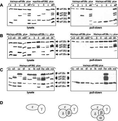
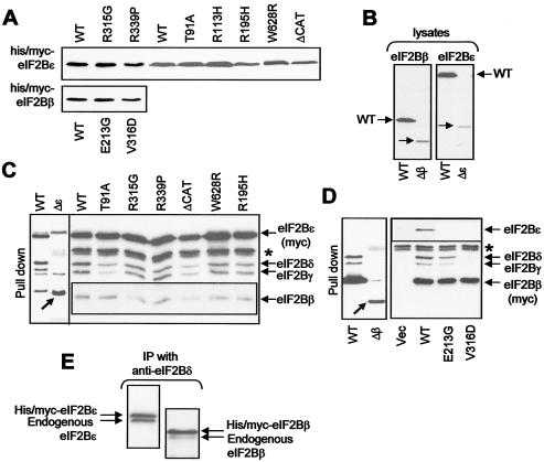
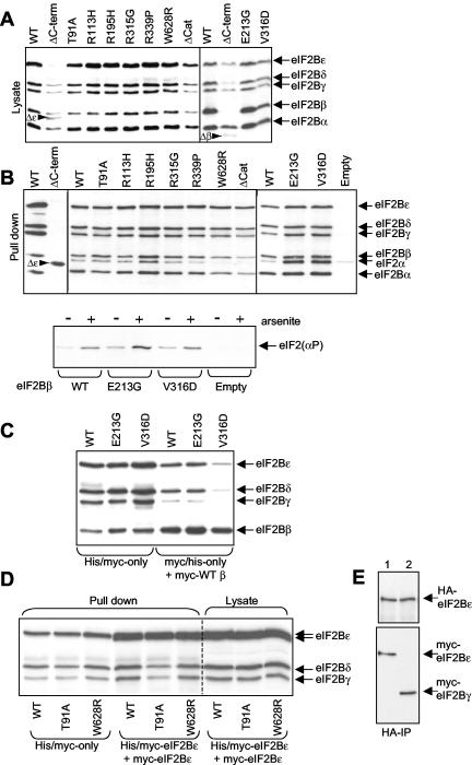
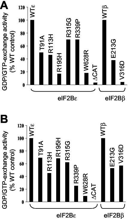
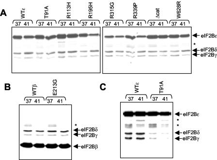
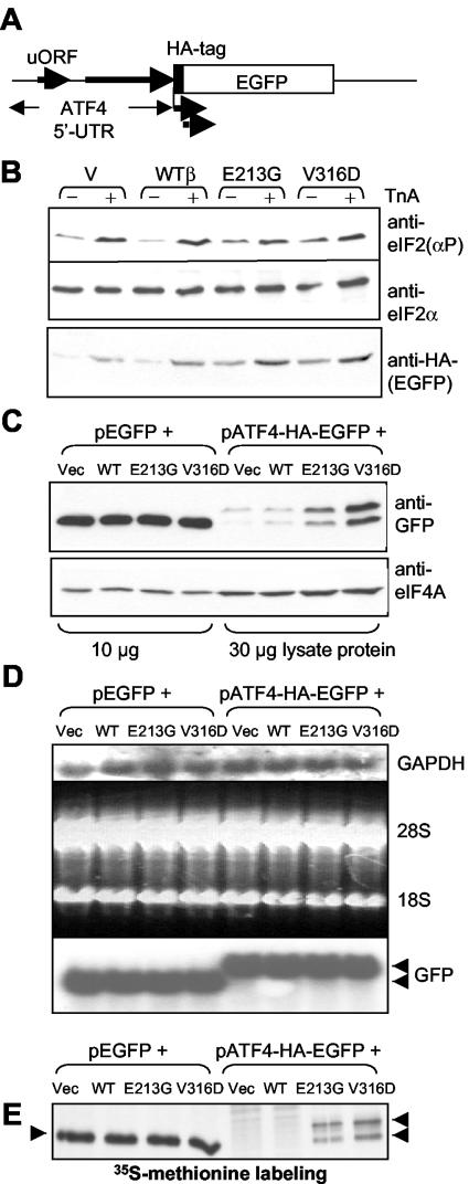
Leukoencephalopathy with vanishing white matter (VWM) is a severe inherited human neurodegenerative disorder that is caused by mutations in the genes for the subunits of eukaryotic initiation factor 2B (eIF2B), a heteropentameric guanine nucleotide exchange factor that regulates both global and mRNA-specific translation. Marked variability is evident in the clinical severity and time course of VWM in patients. Here we have studied the effects of VWM mutations on the function of human eIF2B. All the mutations tested cause partial loss of activity. Frameshift mutations in genes for eIF2Bepsilon or eIF2Bbeta lead to truncated polypeptides that fail to form complexes with the other subunits and are effectively null mutations. Certain point mutations also impair the ability of eIF2Bbeta or -epsilon to form eIF2B holocomplexes and also diminish the intrinsic nucleotide exchange activity of eIF2B. A point mutation in the catalytic domain of eIF2Bepsilon impairs its ability to bind the substrate, while two mutations in eIF2Bbeta actually enhance eIF2 binding. We provide evidence that expression of VWM mutant eIF2B may enhance the translation of specific mRNAs. The variability of the clinical phenotype in VWM may reflect the multiple ways in which VWM mutations affect eIF2B function.


Figures

FIG. 1.
FIG. 1.
Formation of eIF2B complexes in HEK 293 cells. HEK 293 cells were transfected with constructs encoding His-myc-tagged human eIF2Bɛ or -β as indicated, together with vectors encoding myc-tagged versions of the other subunits, as indicated. Samples of cell lysate were either analyzed directly by SDS-PAGE and immunoblotting with anti-myc (lysate) or were first subjected to chromatography on Ni-NTA-agarose prior to this type of analysis (pull-down). Positions of the tagged eIF2B subunits are shown. *, a band that cross-reacts nonspecifically with the anti-myc antibody; all, cells were cotransfected with cDNAs for all four of the other eIF2B subunits. The weak signals in, e.g., the lanes for αδɛ with His-myc-eIF2Bβ in panel C may reflect some “bridging” of the recombinant subunits by the endogenous eIF2B subunits. Panel D presents a cartoon summarizing the data, showing which complexes between eIF2B subunits can form in mammalian cells, i.e., eIF2Bγɛ, eIF2Bβγδɛ, and eIF2Bαβγδɛ.
FIG. 2.
FIG. 2.
Formation of complexes containing a VWM mutant version of eIF2Bβ or ɛ. HEK 293 cells were transfected with vectors for WT or selected VWM mutants of the β or ɛ subunits of eIF2B, as fusions with His and myc tags, or with the empty vector (Vec). (A and B) Samples of cell lysate were analyzed by SDS-PAGE and Western blotting by using anti-myc. (C and D) Samples of cell lysate were subjected to chromatography on Ni-NTA-agarose prior to immunoblot analysis of the bound material with anti-myc, -eIF2Bγ, and -eIF2Bδ as well as anti-eIF2Bβ (C) or -eIF2Bɛ (D). Δɛ and Δβ in panels B and D indicate the frameshift mutants, the positions of the truncated polypeptides that they encode being indicated by diagonal arrows. Positions of the other, endogenous eIF2B subunits are also shown. Weak signals for the anti-eIF2Bβ and ɛ antisera in panels C and D, respectively, mean that their signals are shown for longer exposures as insert panels. The asterisk indicates a nonspecific detection by anti-myc of a protein that binds nonspecifically to Ni-NTA-agarose. (E) Cells were transfected with vectors for myc/His-eIF2Bɛ or -β. Lysates were subjected to IP with anti-eIF2Bδ, and precipitates were analyzed by SDS-PAGE and immunoblotting with anti-eIF2Bɛ (left side) or anti-eIF2Bβ (right side).
FIG. 3.
FIG. 3.
Formation of complexes containing VWM variants of eIF2Bβ or -ɛ. HEK 293 cells were transfected with vectors for WT or selected VWM mutants of the β or ɛ subunits of eIF2B, or the ΔCAT mutant of eIF2Bɛ, as fusions with His and myc tags, and with vectors encoding myc-tagged version of the other four subunits. As a negative control, cells were transfected with empty vectors (indicated). Samples of cell lysate were analyzed directly by SDS-PAGE and Western blotting (A) or first subjected to chromatography on Ni-NTA-agarose prior to analysis by SDS-PAGE-immunoblotting (B). Blots were developed with anti-myc (A and B) and anti-eIF2α (B). Positions of the myc-tagged eIF2B subunits and of eIF2α are shown. Δɛ and Δβ indicate the positions of the truncated polypeptides arising from the frameshift mutations. In the lower part of panel B, only the vectors for WT or mutant eIF2Bβ were used (or empty vector as negative control). Samples of cell lysate were subjected to chromatography on Ni-NTA-agarose prior to analysis by SDS-PAGE and Western blotting with anti-eIF2(Ser51[P]). Equal expression of eIF2B subunits was confirmed by probing with anti-myc (data not shown). Where indicated, cells were pretreated with sodium arsenite (0.5 mM, 30 min) prior to lysis. Cells were also transfected with the empty vector, and similar pull-downs were performed to confirm that very little, if any, eIF2 was retained nonspecifically on the resin (B, upper and lower sections). (C and D) As done for panel B, vectors for the indicated His-myc-tagged eIF2B subunits were used together with myc-tagged versions of the other four WT subunits. Where indicated, cells also received DNA for the myc-tagged versions of eIF2Bɛ or β. Data are immunoblots of material retained on Ni-NTA-agarose (except where indicated, “lysate”). In panel D, the two arrows for eIF2Bɛ indicate that the broad band seen for this subunit actually consists of a poorly resolved doublet of species, the lower one being the myc-tagged eIF2Bɛ and the upper one being the myc/His-tagged subunit. (E) HEK 293 cells were transfected with HA- and myc-tagged versions of eIF2Bɛ; those shown in lane 2 also received the vector for myc-eIF2Bγ. Samples of lysates were subjected to IP with anti-HA followed by SDS-PAGE and immunoblotting, development being with anti-HA (upper section) or anti-myc (lower section).
FIG. 4.
FIG. 4.
Effects of VWM mutations on the activity of eIF2B. HEK 293 cells were transfected with vectors for WT or selected VWM mutant versions of the β or ɛ subunits of eIF2B, or the ΔCAT mutant of eIF2Bɛ, as fusions with His and myc tags (A and B), and with vectors encoding myc-tagged version of the other four subunits (B). Samples of cell lysate were subjected to chromatography on Ni-NTA-agarose prior to analysis. Levels of expression were carefully assessed by SDS-PAGE and Western blot analysis (using anti-myc) of the bound material to normalize the amounts used in each assay. Appropriate amounts of this bound material were then assayed for GDP/GTP exchange activity by using eIF2-[3H]GDP complexes as the substrate. Data are shown as percentage of activity with corresponding WT subunit and are typical of between 4 and 10 experiments performed.
FIG. 5.
FIG. 5.
Analysis of potential thermosensitivity of eIF2B complexes containing WT or VWM mutant eIF2B subunits. HEK 293 cells were transfected with vectors for WT or selected VWM mutant versions of the ɛ (A and C) or β (B) subunits of eIF2B, as fusions with His and myc tags. In some cases, cells were held at 41° for 2 h prior to lysis instead of at the normal temperature of 37°C. Samples of lysate were subjected to chromatography on Ni-NTA-agarose prior to analysis by SDS-PAGE and Western blotting with anti-myc for His-myc-eIF2Bɛ (A and C) and for His-myc-eIF2Bβ (B) and anti- eIF2Bγ and eIF2Bδ. *, nonspecific detection by anti-myc of proteins that bind nonspecifically to Ni-NTA-agarose.
FIG. 6.
FIG. 6.
VWM mutants of eIF2Bβ enhance expression of EGFP from a reporter construct containing the ATF4 5′ UTR. (A) Diagram showing the reporter construct based on the 5′ UTR of the ATF4 mRNA. The broad arrows indicate that the reporter vector gives rise to two translation products, one of which lacks the HA tag (lower, broken arrow) (see panels B, C, and E). (B to E) HEK 293 cells were transfected with vectors for WT eIF2Bβ or the indicated VWM mutants or the empty vector (Vec), along with the reporter construct containing the 5′ UTR of mouse ATF4 ahead of the coding region for HA-tagged EGFP. (C) The control reporter (pEGFP) was used in place of the ATF4-based vector where indicated. In panels B and C, samples were analyzed by SDS-PAGE and Western blotting by using the indicated antisera. (Anti-eIF4A was used as a loading control.) For panel B, where indicated, cells were treated with TnA (2.5 μg/ml) for 16 h prior to lysis. The two bands seen with anti-GFP for the ATF4-based vector in panel C reflect the fact that a second in-frame AUG within the HA-EGFP cistron gives rise to a product lacking the HA tag (only the upper band is recognized by anti-HA). This downstream start codon is likely the start codon for the EGFP itself. This phenomenon probably reflects continued ribosome movement beyond the ATF4 5′ UTR leading up to acquisition by the ribosome of active eIF2, allowing use of the downstream start codon. (D) Samples of lysate were processed for Northern blot analysis. Shown are the autoradiography data obtained with probes for GAPDH (top) and GFP (bottom) and the stained gel showing the positions of the 18S and 28S rRNAs (middle: the top and middle panels serve as loading controls). The ATF4-EGFP mRNA is, as expected, larger than the EGFP one. (E) Cells were labeled with [35S]methionine (see Materials and Methods), and samples of cell lysate were analyzed by IP with anti-GFP, SDS-PAGE, and fluorography to detect the labeled HA-EGFP polypeptides (positions indicated by arrowheads). Again, two bands are seen here, for the reason described above.

References

    1. Asano, K., T. Krishnamoorthy, L. Phan, G. D. Pavitt, and A. G. Hinnebusch. 1999. Conserved bipartite motifs in yeast eIF5 and eIF2Bɛ, GTPase-activating and GDP-GTP exchange factors in translation initiation, mediate binding to their common substrate eIF2. EMBO J. 18:1673-1688. - PMC - PubMed
    1. Bushman, J. L., A. I. Asuru, R. L. Matts, and A. G. Hinnebusch. 1993. Evidence that GCD6 and GCD7, translational regulators of GCN4, are subunits of the guanine nucleotide exchange factor for eIF-2 in Saccharomyces cerevisiae. Mol. Cell. Biol. 13:1920-1932. - PMC - PubMed
    1. Clemens, M. J. 2001. Initiation factor eIF2α phosphorylation in stress responses and apoptosis. Prog. Mol. Subcell. Biol. 27:57-89. - PubMed
    1. Colthurst, D. R., D. G. Campbell, and C. G. Proud. 1987. Structure and regulation of eukaryotic initiation factor eIF-2. Sequence of the site in the alpha subunit phosphorylated by the haem-controlled repressor and by the double-stranded RNA-activated inhibitor. Eur. J. Biochem. 166:357-363. - PubMed
    1. Craddock, B. L., and C. G. Proud. 1996. The α-subunit of mammalian initiation factor eIF-2B is essential for catalytic activity. Biochem. Biophys. Res. Commun. 220:843-847. - PubMed

Publication types

MeSH terms

Substances