. 2004 Apr 13;101(15):5553-8.
doi: 10.1073/pnas.0307575101. Epub 2004 Apr 1.

Designed angiopoietin-1 variant, COMP-Ang1, protects against radiation-induced endothelial cell apoptosis

Affiliations

Designed angiopoietin-1 variant, COMP-Ang1, protects against radiation-induced endothelial cell apoptosis

Chung-Hyun Cho et al. Proc Natl Acad Sci U S A. .

Abstract

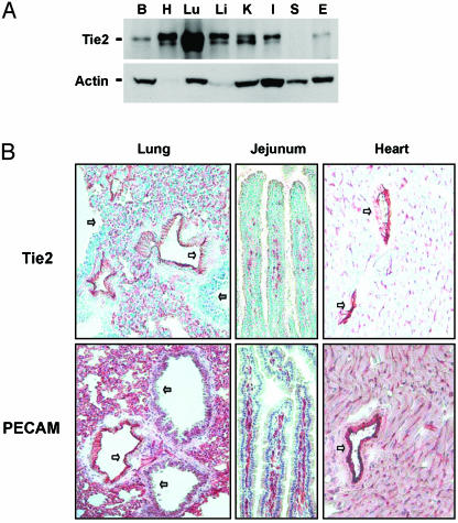
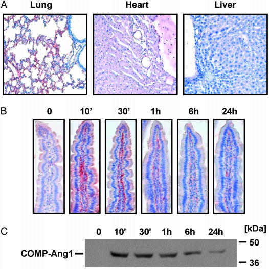
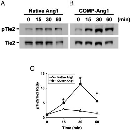
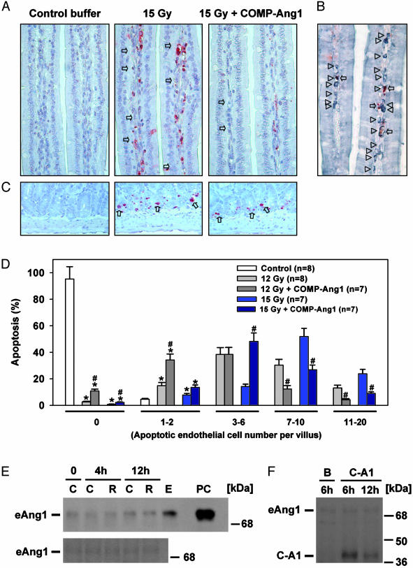
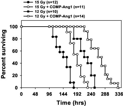
Radiation therapy is a widely used cancer treatment, but it causes side effects even when localized radiotherapy is used. Extensive apoptosis of microvascular endothelial cells of the lamina propria is the primary lesion initiating intestinal radiation damage after abdominal radiation therapy. Many in vitro studies suggest that angiopoietin-1 (Ang1) has potential therapeutic applications in enhancing endothelial cell survival. For in vivo use, we developed a soluble, stable, and potent Ang1 variant, COMP-Ang1. COMP-Ang1 is more potent than native Ang1 in phosphorylating the Tie2 receptor in lung endothelial cells in vivo. Interestingly, COMP-Ang1 administered i.v. was mainly localized to microvascular endothelial cells of the intestinal villi and lung but not to microvascular endothelial cells of the liver. In irradiated mice, i.v. COMP-Ang1 protected against radiation-induced apoptosis in microcapillary endothelial cells of the intestinal villi and prolonged survival. Thus, COMP-Ang1 could be used as a therapeutic protein for specific protection against endothelial cell injury.


Figures

Fig. 1.
Fig. 1.
Distribution and localization of Tie2. (A) Distribution of Tie2 in several organs including brain (B), heart (H), lung (Lu), liver (Li), kidney (K), small intestine (I), spleen (S), and ear (E) of adult mice. Protein lysates from each organ were immunoprecipitated and immunoblotted with anti-Tie2 Ab (Upper). Each protein lysate was also immunoblotted with anti-actin Ab to verify equal amounts of total protein (Lower). (B) Immunohistochemical staining of Tie2 counterstained with methyl green and PECAM-1 counterstained with Meyer's hematoxylin. Most Tie2 staining corresponds to PECAM staining in alveolar capillary endothelial cells (red-brown). Unstained cells are alveolar epithelial cells. White arrows indicate large blood vessels with positive staining. Gray arrows indicate bronchioles with negative staining.
Fig. 2.
Fig. 2.
COMP-Ang1 injected i.v. localizes in microvascular endothelial cells of lung and intestinal villi. (A) Mice were given COMP-Ang1 (30 μg) i.v. and killed after 10 min. The indicated tissues were immunostained with PA-hAng1FD-1 Ab and counterstained with Meyer's hematoxylin. Reddish-brown indicates immunopositive signals. (B) Mice were given COMP-Ang1 (30 μg) i.v. and killed at the indicated times. Intestinal villi were stained as described in A.(C) Part of the jejunum was homogenized, 100 μg of protein homogenates was separated by SDS/PAGE, and the quantity of COMP-Ang1 was measured by immunoblotting with PA-hAng1FD-1 Ab. Results were similar from three independent experiments. Numbers and bars on the right indicate molecular sizes (kDa).
Fig. 3.
Fig. 3.
i.v. injection of COMP-Ang1 induces Tie2 phosphorylation in lung. In vivo phosphorylation of Tie2 stimulated by i.v. injection of native Ang1 (60 μg) (A) or COMP-Ang1 (30 μg) (B) assayed at the indicated times after injection. Tie2 was immunoprecipitated from lung protein lysate and immunoblotted with anti-phosphotyrosine to detect phosphorylated Tie2 (pTie2; Upper). The membrane was stripped and reprobed with anti-Tie2 Ab (Lower) to verify the equal loading of protein in each lane. (C) The relative ratio measured at time 0 is presented arbitrarily as 1. Datapoints represent the mean ± SD from five experiments. *, P < 0.05, compared with native Ang1.
Fig. 4.
Fig. 4.
COMP-Ang1 protects against radiation-induced microvascular apoptosis and death without changes of endogenous Ang1. (A) Mice were given whole-body irradiation with 15 Gy and killed after 4 h. Whereas small intestinal villi from control mice have almost no apoptosis, villi from irradiated mice reveal extensive apoptosis (arrows) in lamina propria cells. Injection of COMP-Ang1 markedly reduced radiation-induced apoptosis in lamina propria cells. (B) Costaining of PECAM-1 (gray-pastel; arrowheads), and deoxynucleotidyl transferase-mediated dUTP nick-end labeling (red-brown; arrows) allows for counting the apoptotic endothelial cells among the lamina propria cells of small-intestinal villi of the irradiated mice. (C) Whereas small intestinal crypts from control mice have almost no apoptosis, crypts from irradiated mice reveal extensive apoptosis (arrows) in the lamina propria. Injection of COMP-Ang1 did not reduce radiation-induced apoptosis in crypt cells. (D) Histograms of percentge of apoptotic endothelial cells in the lamina propria of ≈1,400–1,600 villi from each group (200 villi per mouse). Small-intestinal specimens were obtained at 4 h after 12 or 15 Gy irradiation with or without i.v. injection of COMP-Ang1. Apoptotic endothelial cells were counted in each villus, categorized (0, 1–2, 3–6, 7–10, or 11–20), and calculated as a percentage with 200 villi. Bars represent the mean ± SD. from each group. *, P < 0.05 versus control in 0 or 1–2.#, P < 0.05 versus 12 or 15 Gy irradiation only. (E) Mice were given whole-body irradiation with 15 Gy and killed at 4 h and 12 h. Part of the jejunum (Upper) and lung (Lower) was homogenized, 200 μg of protein homogenates was separated by SDS/PAGE, and the relative amount of endogenous Ang1 (eAng1) was determined by immunoblotting. Results were similar from three independent experiments. C, control; R, irradiation; E, 100 μg of protein homogenates from mouse embryonic day 12; PC, 20 ng of recombinant mouse native Ang1 as a positive control. Numbers and bars on the right indicate molecular masses (kDa). (F) Mice were given whole-body irradiation with 15 Gy and killed after 6 h or 24 h. The mice were injected i.v. with 100 μg of COMP-Ang1 (C-A1) in 33-μg doses delivered 30 min before, 30 min after, and 90 min after irradiation. For control mice, 100 μg of BSA (B) was injected at the same time points. Part of the jejunum was homogenized, 200 μg of protein homogenates were separated by SDS/PAGE, and the relative amount of COMP-Ang1 and endogenous Ang1 (eAng1) was determined by immunoblotting. Results were similar from three independent experiments. B, BSA at 6 h. Numbers and bars on the right indicate molecular masses (kDa).
Fig. 5.
Fig. 5.
Treatment with COMP-Ang1 prolongs survival in mice irradiated with 12 or 15 Gy. Survival was monitored every 12 h after irradiation. Numbers in parentheses indicate the number of animals per group. Two-tailed Fisher's exact test was performed to compare control and COMP-Ang1-treated mice (P = 0.002 for 12 Gy; P = 0.003 for 15 Gy).

References

    1. Paris, F., Fuks, Z., Kang, A., Capodieci, P., Juan, G., Ehleiter, D., Haimovitz-Friedman, A., Cordon-Cardo, C. & Kolesnick, R. (2001) Science 293, 293–297. - PubMed
    1. Okunieff, P., Mester, M., Wang, J., Maddox, T., Gong, X., Tang, D., Coffee, M. & Ding, I. (1998) Radiat. Res. 150, 204–211. - PubMed
    1. Veikkola, T. & Alitalo, K. (1999) Semin. Cancer Biol. 9, 211–220. - PubMed
    1. Davis, S., Aldrich, T. H., Jones, P. F., Acheson, A., Compton, D. L., Jain, V., Ryan, T. E., Bruno, J., Radziejewski, C., Maisonpierre, P. C., et al. (1996) Cell 87, 1161–1169. - PubMed
    1. Yancopoulos, G. D., Davis, S., Gale, N. W., Rudge, J. S., Wiegand, S. J. & Holash, J. (2000) Nature 407, 242–248. - PubMed

Publication types

MeSH terms

LinkOut - more resources