Comparative Study
. 2004 May;74(5):1064-73.
doi: 10.1086/420795. Epub 2004 Apr 1.

Identification of a novel gene (HSN2) causing hereditary sensory and autonomic neuropathy type II through the Study of Canadian Genetic Isolates

Affiliations
Comparative Study

Identification of a novel gene (HSN2) causing hereditary sensory and autonomic neuropathy type II through the Study of Canadian Genetic Isolates

Ronald G Lafreniere et al. Am J Hum Genet. 2004 May.

Abstract

Hereditary sensory and autonomic neuropathy (HSAN) type II is an autosomal recessive disorder characterized by impairment of pain, temperature, and touch sensation owing to reduction or absence of peripheral sensory neurons. We identified two large pedigrees segregating the disorder in an isolated population living in Newfoundland and performed a 5-cM genome scan. Linkage analysis identified a locus mapping to 12p13.33 with a maximum LOD score of 8.4. Haplotype sharing defined a candidate interval of 1.06 Mb containing all or part of seven annotated genes, sequencing of which failed to detect causative mutations. Comparative genomics revealed a conserved ORF corresponding to a novel gene in which we found three different truncating mutations among five families including patients from rural Quebec and Nova Scotia. This gene, termed "HSN2," consists of a single exon located within intron 8 of the PRKWNK1 gene and is transcribed from the same strand. The HSN2 protein may play a role in the development and/or maintenance of peripheral sensory neurons or their supporting Schwann cells.


Figures
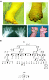
Figure  1
Figure 1
Phenotype of HSAN type II and pedigrees for large Newfoundland families described in this study. a, Examples of clinical presentations. Patients in these families suffer from loss of touch and pain sensation leading to ulceration, infection, muscle atrophy, and amputation of digits. b, Partial pedigrees of families F1 and F2. Squares and circles represent males and females, respectively. Blackened symbols indicate individuals with HSAN type II. Unblackened symbols indicate individuals normal by clinical examination, and symbols with a question mark indicate individuals who have not been examined. Diagonal slashes indicate deceased individuals. All examined individuals were included in the genome scan or the subsequent fine mapping.
Figure  2
Figure 2
Haplotype analysis and candidate region. a, Haplotypes were constructed for all genotyped individuals in families F1 and F2, but here only affected haplotypes are shown. GENEHUNTER v2.1_r2 beta (Kruglyak et al. ; Kruglyak and Lander 1998) was used to construct haplotypes on pedigree sections, which were then manually combined. Haplotypes are designated with pedigree (F) number, individual number, and chromosome (a or b) number. The three different haplotype groups bearing causal mutations are numbered 1–3. Allele sizes are given in bp. Markers are indicated at the top, with their physical distances from the telomere given in kb on the basis of the July 2003 build 34 human genome assembly and their genetic positions, according to the deCODE map. Portions of haplotypes encompassing the HSN2 locus are boxed. Haplotypes for F1 and F2 are phased, except for F1–122/124, which was inferred from genotypes in parent F1-122 and deceased offspring F1-124. Phasing of F3-301 and F3-302 was inferred on the basis of allele sharing with a region of homozygosity in F4-301. Recombination breakpoints in the shared haplotype in individuals F1-171 and F1-124 placed the HSAN II locus between markers CA1AC0021054 and D12S1642. Affected French Canadian samples were subsequently genotyped for markers in this region. A recombination event in one of the sisters in F3 positioned the centromeric boundary at marker CA1AC005183. b, Physical map of the HSAN II candidate region, showing markers used in the genetic analysis and candidate genes represented as unblackened arrows (derived from UCSC genome annotation). The arrows indicate the direction of transcription for each gene. The distances are given in kb from the telomere on the basis of the July 2003 build 34 human genome assembly. The HSN2 gene is indicated by a blackened arrow. To identify unannotated conserved ORFs, the human genomic sequence representing the entire HSAN II candidate interval (∼1.0 Mb) was downloaded with case toggled to highlight the mouse translated BLAT track (which represents regions showing significant protein homology between human and mouse). These sequences were assembled into a contig, to which all exons of previously identified candidate genes were added. All sequences were manipulated using DNASTAR software. The contig was scanned for novel conserved fragments (>80% conservation over >100 bp). This identified 64 novel fragments, which were then tested for functional homology using (1) BLASTn against the nr database, (2) BLASTn against the dbEST, and (3) BLASTx against the nr database available from the NCBI Web site. The HSN2 gene was, by a substantial margin, the most highly conserved and the longest ORF among these fragments. It should be noted that various gene-prediction algorithms—such as ECgene, Geneid, SGP, and Twinscan—also predict the HSN2 ORF, in whole or in part, either as a separate exon or as a potential alternative exon of the PRKWNK1 gene (for which there is no evidence among mammalian ESTs).
Figure  3
Figure 3
Mutations in the HSN2 gene in affected individuals. To screen the HSN2 ORF for mutations, three separate amplicons were designed (for primers, see appendix A [online only]) using PrimerSelect (DNASTAR) and were purchased from BioCorp. Amplified products were sequenced at the Montreal Genome Centre sequencing facility. Sequence traces were aligned using SeqManII (DNASTAR) and were inspected visually for mutations. Each panel displays a sequencing trace from a normal control (above) and from an affected individual (below) according to the following: a, Homozygous c.594delA mutation in patient F1-70 from Newfoundland, causing a frameshift in codon 198 leading to premature truncation to a 206-aa peptide. This mutation cosegregated perfectly with the disease in the F1 and F2 families from Newfoundland as predicted from the haplotype analysis. b, Homozygous c.918–919insA mutation in patient F5-301 from Nova Scotia, causing a frameshift in codon 307 and truncation to a 318-aa peptide. Both Nova Scotia siblings shared this mutation, which was also heterozygous in the French Canadian F3 samples. c, Homozygous c.943C→T nonsense mutation in French Canadian patient F4-301, changing codon 315 (CAG, encoding glutamine) to a TAG stop codon, and truncating the protein to 314 aa. This mutation was also heterozygous in the F3 samples. All sequence traces are from the forward strand. To genotype each of the three mutations in additional family members and population controls, we used PCR-RFLP or capillary electrophoresis analysis (details of mutation detection sequencing and genotyping in appendix A [online only]).
Figure  4
Figure 4
Alignment of predicted HSN2 peptide sequences for vertebrate orthologs. a, The conserved HSN2 ORF was identified from the genomic assemblies of human, mouse, rat, and zebrafish (Zfish) and from the pig cDNA clone. BLAST searches were done through the NCBI Web site. Genomic sequences for HSN2 orthologs were identified within GenBank sequence files AC004765 (human), AC106932 and AC106348 (rat), AC113092 (mouse), and BX321885 (zebrafish). Prosite searches were done through the EBI server. For the predicted peptide sequence alignment, sequences were aligned using Clustal 1.8, available from the BCM Search Launcher Web site. Aligned sequences then were shaded using BOXSHADE. Functional bioinformatics analysis was performed using SignalP. Conserved residues are shaded. Residues are numbered from the most upstream start ATG. Asterisks (*) above N-terminal residues indicate potential initiating methionines; asterisks at the C-termini indicate the stop codons. The signal peptide and predicted cleavage residue are indicated at the N-terminus. Positions of causative mutations in human populations are indicated. b, Alignment of 3′ ends of resequenced human and pig cDNA clones with the human genomic sequence, showing conservation of polyadenylation sites. The putative polyadenylation signal is shown above the aligned sequences.

References

Electronic-Database Information

    1. BCM, http://searchlauncher.bcm.tmc.edu/multi-align/multi-align.html
    1. BOXSHADE, http://www.ch.embnet.org/software/BOX_form.html
    1. EBI, http://www.ebi.ac.uk/ppsearch/
    1. Gene Discovery Program for Functional Genomics in Pig Reproduction, http://pigest.genome.iastate.edu/
    1. HUGO Gene Nomenclature Committee, http://www.gene.ucl.ac.uk/nomenclature/

References

    1. Anderson SL, Coli R, Daly IW, Kichula EA, Rork MJ, Volpi SA, Ekstein J, Rubin BY (2001) Familial dysautonomia is caused by mutations of the IKAP gene. Am J Hum Genet 68:753–758 - PMC - PubMed
    1. Axelrod FB (2002) Hereditary sensory and autonomic neuropathies: familial dysautonomia and other HSANs. Clin Auton Res Suppl 12:2–1410.1007/s102860200014 - DOI - PubMed
    1. Basu S, Paul DK, Basu S (2002) Four siblings with type II hereditary sensory and autonomic neuropathy. Indian Pediatr 39:870–874 - PubMed
    1. Bejaoui K, Wu C, Scheffler MD, Haan G, Ashby P, Wu L, de Jong P, Brown RH Jr. (2001) SPTLC1 is mutated in hereditary sensory neuropathy, type I. Nat Genet 27: 261–26210.1038/85817 - DOI - PubMed
    1. Choate KA, Kahle KT, Wilson FH, Nelson-Williams C, Lifton RP (2003) WNK1, a kinase mutated in inherited hypertension with hyperkalemia, localizes to diverse Cl- -transporting epithelia. Proc Natl Acad Sci USA 100:663–66810.1073/pnas.242728499 - DOI - PMC - PubMed

Publication types

MeSH terms

Associated data