. 2004;31(1):61-8.

The surgical anatomy of experimental and clinical thoracic organ transplantation

Affiliations

The surgical anatomy of experimental and clinical thoracic organ transplantation

David K C Cooper. Tex Heart Inst J. 2004.

Abstract

The experimental investigation of heart transplantation began almost 100 years ago, but it was not until the studies at Stanford Medical School in the late 1950s and early 1960s that clinical transplantation became a realistic possibility. Barnard performed the 1st human-to-human orthotopic heart transplantation in 1967 and followed this by introducing the technique of heterotopic heart transplantation in 1974. Reitz and colleagues at Stanford performed the 1st successful clinical transplantation of the heart and both lungs in 1981. Two years later, at the Toronto General Hospital, successful single-lung transplantation was performed, followed by bilateral lung transplantation in 1986. Aspects of the surgical techniques of these various experimental and clinical procedures are discussed.


Figures

None
Fig. 1 Experimental heterotopic heart transplantation in the neck. The donor coronary arteries are perfused with oxygenated blood from the recipient's carotid artery. Coronary venous return is ejected through the donor pulmonary artery into the internal jugular vein of the recipient. (The donor heart can be placed in the abdomen with anastomoses to the recipient aorta and inferior vena cava.) AO = aorta; CCA = common carotid artery; IJV = internal jugular vein; LA = left atrium; LV = left ventricle; PA = pulmonary artery; RA = right atrium; RV = right ventricle (From: Cooper DKC, et al., © 1996. Reproduced with kind permission of Kluwer Academic Publishers.)
None
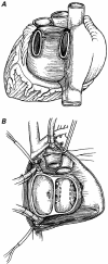
Fig. 2 Orthotopic heart transplantation. A) The recipient ventricles with all 4 valves have been excised. Note the incision in the donor right atrium extending into the atrial appendage to avoid the sinoatrial node. The 1st suture line between the donor and recipient hearts (between the free walls of the left atria) has been started. This will be followed by anastomoses of the 2 atrial septa, the free walls of the right atria, the aortae, and pulmonary arteries, not necessarily in that order. B) The completed operation. IVC = inferior vena cava; SVC = superior vena cava; other abbreviations as for Fig. 1 (From: Cooper DKC, et al., © 1996. Reproduced with kind permission of Kluwer Academic Publishers.)
None
Fig. 3 “Total” orthotopic heart transplantation. A) Posterior aspect of the donor heart: the superior and inferior venae cavae have been retained with the right atrium; the posterior wall of the left atrium is intact except for the orifices of the paired pulmonary veins. B) The recipient pericardial cavity after excision of the native ventricles: the atria will be excised, and the only remaining structures will be the 2 venae cavae and the 2 left atrial cuffs around the pulmonary vein orifices (indicated by dotted lines). (From: Cooper DKC, et al., © 1996. Reproduced with kind permission of Kluwer Academic Publishers.)
None
Fig. 4 Heterotopic heart transplantation. A) The completed operation of left heart support with a heterotopic heart transplant: the respective donor and recipient atria are anastomosed, followed by an end-to-side aortic anastomosis; the coronary venous drainage of the donor heart is drained through the donor pulmonary artery into the recipient right atrium. B) The completed operation of biventricular support: the anastomoses are as above, except that the right ventricular output is directed through the donor pulmonary artery into the recipient pulmonary artery, usually through a synthetic conduit. C) Replacement of the native heart in a patient with a previous heterotopic heart transplant: the shaded areas represent all that remains of the patient's native tissues. D1 = initial heterotopic donor heart; D2 = subsequent orthotopic donor heart; other abbreviations as for Figs. 1 and 2. (From: Cooper DKC, et al., © 1996. Reproduced with kind permission of Kluwer Academic Publishers.)
None
Fig. 5 Transplantation of the heart and both lungs. A) The recipient thorax after excision of the native heart and lungs: the trachea will be divided just above the carina. B) The donor right lung is passed posterior to the remnant of the recipient's right atrium and right phrenic nerve. The donor left lung is passed posterior to the left phrenic nerve. C) The donor right and left lungs have been placed in the respective pleural cavities. The tracheal anastomosis has been completed; the areolar tissue around the donor left atrium can be used to cover the tracheal suture line. The right atrial anastomosis is in progress. Anastomosis of the 2 aortae will complete the operation. AO = aorta; LV = left ventricle; PA = pulmonary artery; RA = right atrium; RV = right ventricle (Figs. 5B and 5C are from: Cooper DKC, et al., © 1996. Reproduced with kind permission of Kluwer Academic Publishers.)
None
Fig. 6 Posterior view of heart, bronchi, and trachea, indicating the collateral blood supply to the bronchi and trachea from coronary arteries. LV = left ventricle (From: Cooper DKC, et al., © 1996. Reproduced with kind permission of Kluwer Academic Publishers.)
None
Fig. 7 Single-lung transplantation. Insertion of the donor lung requires anastomoses between the donor and recipient bronchi, pulmonary arteries, and left atrial cuffs. (From: Cooper DKC, et al., © 1996. Reproduced with kind permission of Kluwer Academic Publishers.)

Similar articles

References

    1. Cooper DKC. Experimental development of cardiac transplantation. Br Med J 1968;4:174–81. - PMC - PubMed
    1. Cooper DKC, Miller LW, Patterson GA. The transplantation and replacement of thoracic organs. The present status of biological and mechanical replacement of the heart and lungs. 2nd ed. Dordrecht: Kluwer Academic Publishers; 1996.
    1. Cass MH, Brock R. Heart excision and replacement. Guys Hosp Rep 1959;108:285–90. - PubMed
    1. Cooper DKC, Wicomb WN, Barnard CN. Storage of the donor heart by a portable hypothermic perfusion system: experimental development and clinical experience. Heart Transplant 1983;2:104–10.
    1. Barnard CN. The operation. A human cardiac transplant: an interim report of a successful operation performed at Groote Schuur Hospital, Cape Town. S Afr Med J 1967;41:1271–4. - PubMed

Publication types

MeSH terms