. 2004 Jun;15(6):2834-41.
doi: 10.1091/mbc.e04-01-0059. Epub 2004 Apr 2.

The role of osteoprotegerin and tumor necrosis factor-related apoptosis-inducing ligand in human microvascular endothelial cell survival

Affiliations

The role of osteoprotegerin and tumor necrosis factor-related apoptosis-inducing ligand in human microvascular endothelial cell survival

L B Pritzker et al. Mol Biol Cell. 2004 Jun.

Abstract

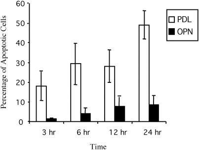
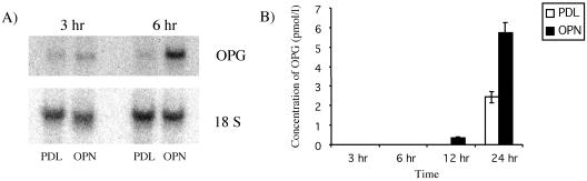
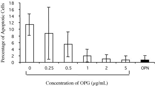
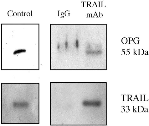
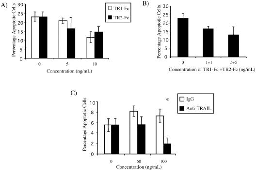
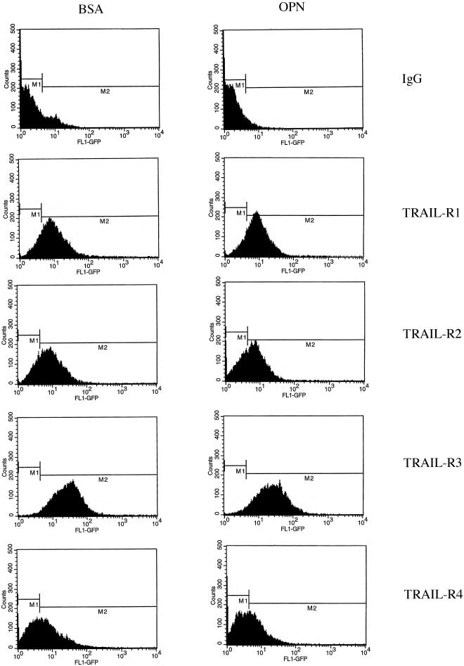
Endothelial cell survival and antiapoptotic pathways, including those stimulated by extracellular matrix, are critical regulators of vasculogenesis, angiogenesis, endothelial repair, and shear-stress-induced endothelial activation. One of these pathways is mediated by alpha(v)beta(3) integrin ligation, downstream activation of nuclear factor-kappaB, and subsequent up-regulation of osteoprotegerin (OPG). In this study, the mechanism by which OPG protects endothelial cells from death was examined. Serum-starved human microvascular endothelial cells (HMECs) plated on the alpha(v)beta(3) ligand osteopontin were protected from cell death. Immunoprecipitation experiments indicated that OPG formed a complex with tumor necrosis factor-related apoptosis-inducing ligand (TRAIL) in HMECs under these conditions. Furthermore, inhibitors of TRAIL, including recombinant soluble TRAIL receptors and a neutralizing antibody against TRAIL, blocked apoptosis of serum-starved HMECs plated on the nonintegrin attachment factor poly-d-lysine. Whereas TRAIL was unable to induce apoptosis in HMECs plated on osteopontin, the addition of recombinant TRAIL did increase the percentage of apoptotic HMECs plated on poly-d-lysine. This evidence indicates that OPG blocks endothelial cell apoptosis through binding TRAIL and preventing its interaction with death-inducing TRAIL-receptors


Figures

Figure 1.
Figure 1.
HMEC survival is increased in cells plated on OPN compared with PDL. HMECs were plated on either OPN or PDL in the absence of serum for 3, 6, 12, or 24 h. Nuclei were then stained with Hoechst dye, and the number of apoptotic cells was determined in three fields at 40×. The percentage of apoptotic cells was then calculated. Cells were considered to be apoptotic on the basis of nuclear condensation and fragmentation. The experiment was repeated three times, and error bars indicate standard deviations for triplicate determinations (analysis of variance, p < 0.0001).
Figure 2.
Figure 2.
OPG RNA and secretion into the media are increased in HMECs plated on OPN. (A) HMECs were plated on either OPN or PDL for 3 and 6 h and the RNA isolated. The samples were run on an agarose gel, and Northern blot analysis was performed. OPG RNA from HMECs plated on OPN was increased at 3 h and was further increased at 6 h. (B) HMECs were plated on either OPN or PDL for 3, 6, 12, or 24 h, and the media were collected and stored at -20°C. The media were concentrated 20-fold by using Microcon filters, and the amount of OPG was quantitated using a sandwich ELISA. A twofold increase in OPG was found in HMECs plated on OPN at 12 and 24 h. Results are representative of three independent experiments; error bars represent standard deviations (analysis of variance, p < 0.0001; three replicates).
Figure 3.
Figure 3.
Recombinant OPG-Fc inhibits apoptosis of HMECs. HMECs were plated on PDL for 10 h in the presence of OPG-Fc concentrations ranging from 0 to 5 μg/ml and on OPN. Nuclei were stained with Hoechst dye and counted in three fields at 40×. The percentage of apoptotic nuclei was then determined. The experiment was repeated three times, and error bars indicate standard deviations (analysis of variance, p < 0.0001; three replicates).
Figure 4.
Figure 4.
TRAIL and OPG coimmunoprecipitate. HMECs were plated on OPN for 16 h, and the lysates were collected in an IP buffer. Mouse IgG and a mAb against TRAIL were each cross-linked to agarose, and each conjugate was then incubated with the cell lysates at 4°C overnight. The bound proteins were eluted off the agarose gel and analyzed by Western blot. Both TRAIL and OPG were detected in the eluted fractions. Recombinant TRAIL and OPG were loaded as positive controls. This result indicated that TRAIL and OPG form a complex that can be immunoprecipitated using an anti-TRAIL antibody. Results are representative of three independent experiments.
Figure 5.
Figure 5.
Soluble TRAIL receptors and neutralizing antibody against TRAIL promotes HMEC survival. (A) HMECs were plated on OPN or PDL for 18 h. TR1-Fc and TR2-Fc were added at concentrations of 0, 5, and 10 ng/ml to cells plated on PDL. Both TR1-Fc and TR2-Fc inhibited serum withdrawal-induced apoptosis (analysis of variance, p < 0.003; three replicates). (B) Treatment of HMECs with a combination of TR1-Fc and TR2-Fc did not result in increased inhibition. (C) Similarly, HMECs were plated on PDL in the presence of a neutralizing monoclonal anti-TRAIL at concentrations of 50 and 100 ng/ml. The percentage of apoptotic cells was determined at 16 h. Treatment of cells with anti-TRAIL resulted in a decrease in the percentage of apoptotic cells. At 100 ng/ml, a threefold difference was seen between IgG and antibody-treated cells (*, Fisher's protected least significant difference, p < 0.02). Both soluble TRAIL receptors and an anti-TRAIL mAb inhibited endothelial cell apoptosis. The experiments were repeated three times, and error bars indicate standard deviations.
Figure 6.
Figure 6.
TRAIL enhances apoptosis of HMECs plated on PDL. HMECs were plated on PDL and recombinant TRAIL was added 30 min later at a concentration of 200 ng/ml. The percentage of apoptotic cells was determined at 3, 6, 10, and 24 h. Treatment of cells plated on PDL with TRAIL resulted in a two- to threefold increase in the percentage of apoptotic cells. HMECs also were plated on OPN and on PDL in the absence of TRAIL. TRAIL has no effect on the apoptotic rate of HMEC plated on OPN (our unpublished data). The results shown are representative of three independent experiments; error bars indicate standard deviations for triplicate determinations (analysis of variance, p < 0.0001).
Figure 7.
Figure 7.
TRAIL-receptor expression on the cell surface is unchanged in HMECs plated on BSA and OPN. The expression of TRAIL-R on the cell surface was examined under conditions of survival (OPN) and apoptosis (cells kept in suspension by plating on BSA). HMECs were plated on OPN or BSA for 4 h and resuspended in a buffer containing 10 μg/ml goat IgG or 10 μg/ml antibody against one of TRAIL-R1, TRAIL-R2, TRAIL-R3, or TRAIL-R4. The cells were then incubated with a FITC-labeled anti-goat antibody and analyzed by flow cytometry. No changes were found in the expression of TRAIL-R between HMECs plated on BSA and those plated on OPN. Results are representative of three independent experiments.

References

    1. Alon, T., Hemo, I., Itin, A., Pe'er, J., Stone, J., and Keshet, E. (1995). Vascular endothelial growth factor acts as a survival factor for newly formed retinal vessels and has implications for retinopathy of prematurity. Nat. Med. 1, 1024-1028. - PubMed
    1. Aoudjit, F., and Vuori, K. (2001). Matrix attachment regulates Fas-induced apoptosis in endothelial cells: a role for c-Flip and implications for anoikis. J. Cell Biol. 152, 633-643. - PMC - PubMed
    1. Brooks, P.C., Clark, R.A., and Cheresh, D.A. (1994a). Requirement of vascular integrin avb3 for angiogenesis. Science 264, 569-571. - PubMed
    1. Brooks, P.C., Montgomery, A.M.P., Rosenfeld, M., Reisfeld, R.A., Hu, T., Klier, G., and Cheresh, D.A. (1994b). Integrin avb3 antagonists promote tumor regression by inducing apoptosis of angiogenic blood vessels. Cell 79, 1157-1164. - PubMed
    1. Bucay, N., et al. (1998). Osteoprotegerin-deficient mice develop early onset osteoporosis and arterial calcification. Genes Dev. 12, 1260-1268. - PMC - PubMed

MeSH terms

Substances