. 2004 Jun 17;23(28):4839-46.
doi: 10.1038/sj.onc.1207634.

Phosphorylation and ubiquitination of oncogenic mutants of beta-catenin containing substitutions at Asp32

Affiliations

Phosphorylation and ubiquitination of oncogenic mutants of beta-catenin containing substitutions at Asp32

Mohamed Al-Fageeh et al. Oncogene. .

Abstract

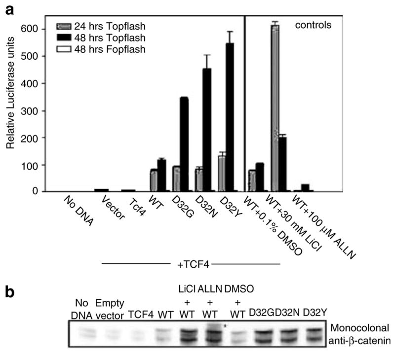
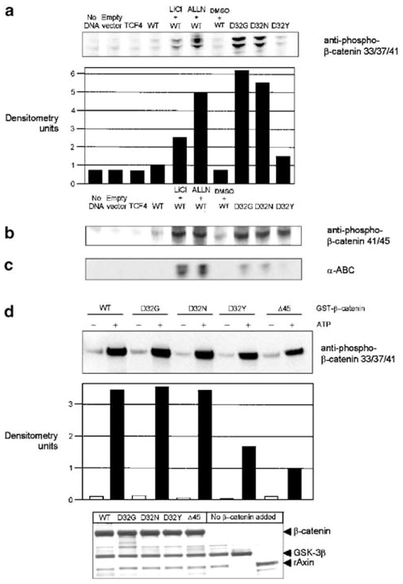
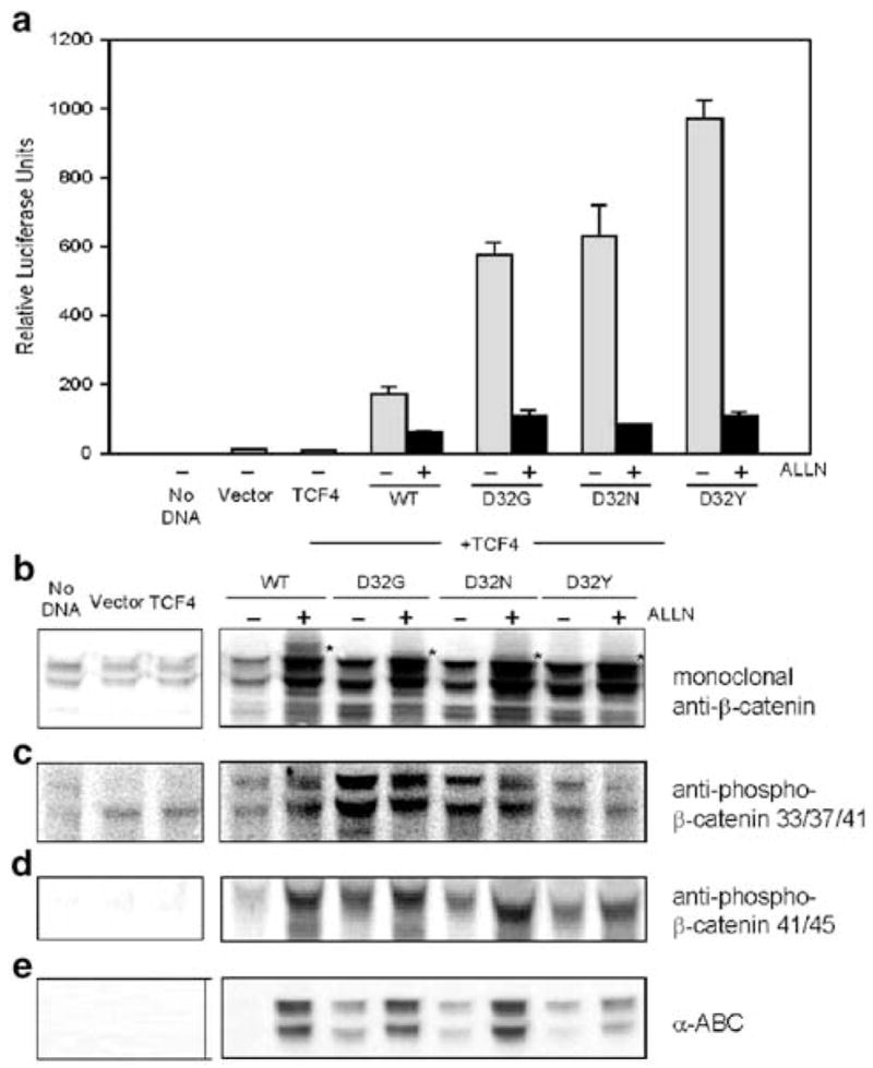
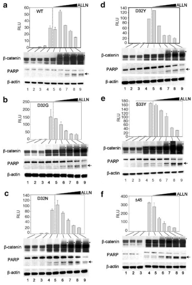
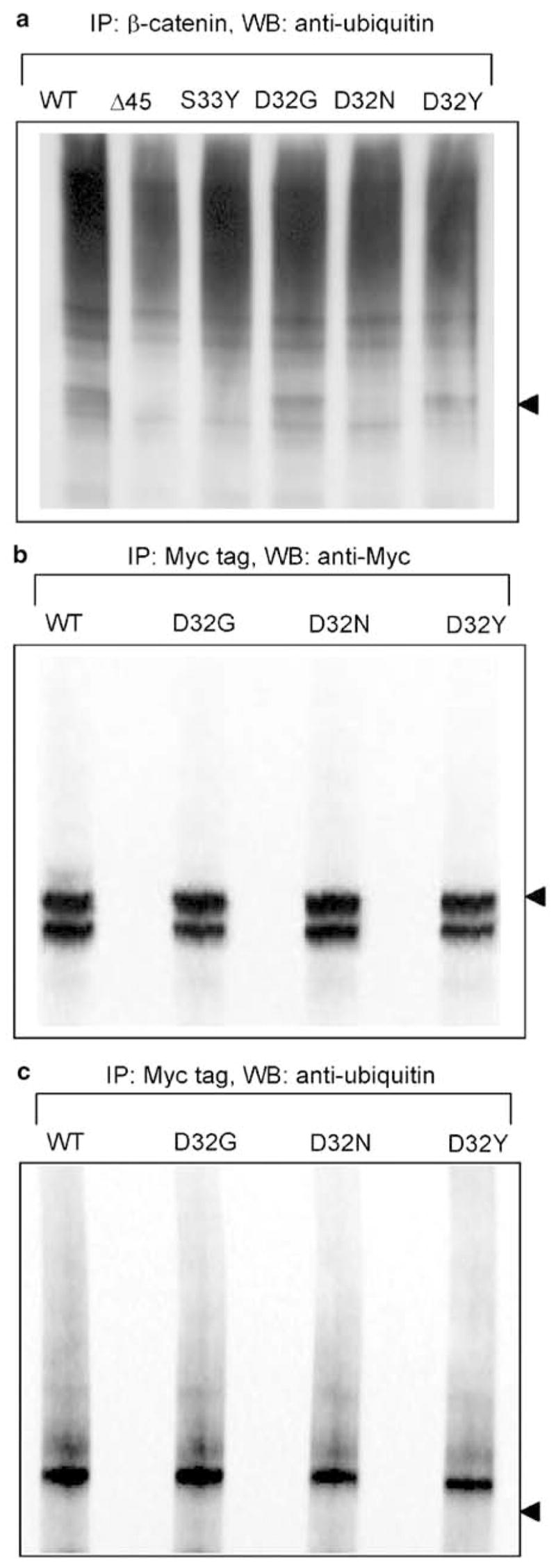
Beta-Catenin, a member of the Wnt signaling pathway, is downregulated by glycogen synthase kinase-3beta (GSK-3beta)-dependent phosphorylation of Ser/Thr residues in the N-terminus of the protein, followed by ubiquitination and proteosomal degradation. In human and rodent cancers, mutations that substitute one of the critical Ser/Thr residues in the GSK-3beta region of beta-catenin stabilize the protein and activate beta-catenin/TCF/LEF target genes. This study examined three oncogenic beta-catenin mutants from rat colon tumors containing substitutions adjacent to amino-acid residue Ser33, a key target for phosphorylation by GSK-3beta. Compared with wild-type beta-catenin (WT), the beta-catenin mutants D32G, D32N, and D32Y strongly activated TCF-4-dependent transcription in HEK293 cells, and there was accumulation of beta-catenin in the cell lysates. Immunoblotting with phosphospecific antibodies indicated that there was little if any effect on the phosphorylation of Ser37, Thr41 or Ser45; however, the phosphorylation of Ser33 appeared to be affected in the beta-catenin mutants. Specifically, antiphospho-beta-catenin 33/37/41 antibody identified high, intermediate and low expression levels of phosphorylated beta-catenin in cells transfected with D32G, D32N and D32Y, respectively. Experiments with the proteosome inhibitor N-acetyl-Leu-Leu-norleucinal (ALLN) revealed ubiquitinated bands on all three mutant beta-catenins, as well as on WT beta-catenin. The relative order of ubiquitination was WT>D32G>D32N>D32Y, in parallel with findings from the phosphorylation studies. These results are discussed in the context of previous studies, which indicated that amino-acid residue D32 lies within the ubiquitination recognition motif of beta-catenin.


Figures

Figure 1
Figure 1
Activation of TCF-4-dependent transcription by WT and mutant β-catenins. (a) A plasmid expressing a TCF/LEF-responsive reporter, TOPflash, or the negative reporter FOPflash (see Morin et al., 1997), was cotransfected into HEK293 cells with empty vector (vector), a plasmid expressing TCF-4 or TCF-4 vector plus a plasmid expressing WT or mutant (D32G, D32N, D32Y) β-catenins. Reporter activation was assessed as luciferase activity in aliquots of cell lysates normalized for protein content. The GSK-3β inhibitor LiCl (30 mM) and the proteosome inhibitor N-acetyl-Leu-Leu-norleucinal (ALLN, 100 μM) were included as controls. The data are given as mean ± s.d. of triplicates. (b) Cell lysates obtained 48 h post-transfection were subjected to immuno-blot analysis with monoclonal anti-β-catenin antibody, as well as antibody to β-actin (not shown). The asterisk denotes the position of high molecular weight band(s), detected after treatment with ALLN but not LiCl
Figure 2
Figure 2
Immunodetection of β-catenin using phosphospecific antibodies. The cell lysates described in Figure 1b were probed with the following antibodies: (a) antiphospho-β-catenin 33/37/41, (b) antiphospho-β-catenin 41/45 and (c) α-ABC (α-active β-catenin, for β-catenin unphosphorylated at Ser45 and Thr41). (d) An in vitro kinase assay was performed with purified GST-tagged rAxin, GSK-3β and WT or mutant β-catenins, in the presence and absence of ATP. Reaction products were analysed by SDS–PAGE with immunodetection using anti-pβ-catenin-33/37/41 antibody. Equal protein loading was confirmed by silver staining (bottom); the last three lanes, respectively, were controls lacking β-catenin but containing GSK-3β plus rAxin, GSK-3β alone or rAxin alone
Figure 3
Figure 3
Effect of ALLN on β-catenin/TCF-4-dependent reporter activity and β-catenin expression in HEK293 cells. (a) Reporter activities (TOPflash) were assessed 48 h post-transfection, as described in Figure 1a. Data shown in the figure (mean ± s.d., n =3) are representative findings from three separate experiments conducted in the presence and absence of 100 μM ALLN. The corresponding cell lysates were immunoblotted with antibodies to (b) total β-catenin (monoclonal anti-β-catenin), (c) antiphospho-β-catenin 33/37/41, (d) antiphospho-β-catenin 41/45 and (e) α-ABC. In (b), the asterisk denotes the position of higher molecular band(s), seen after treatment with ALLN
Figure 4
Figure 4
Dose–response for inhibition by ALLN of β-catenin/TCF-4-dependent reporter activity in HEK293 cells, and the expression of β-catenin and poly(ADP-ribose) polymerase (PARP). Cells were transfected with (a) WT, (b) D32G, (c) D32N, (d) D32Y, (e) S33Y or (f) Δ45 β-catenin. TOPflash reporter activities (mean ± s.d., n =3) were determined as described in Figure 3a –lane 1, no DNA; lane 2, empty vector; lane 3, TCF-4; lane 4, TCF-4 plus β-catenin; lanes 5–9, same as lane 4, but with 0 (DMSO vehicle alone), 5, 10, 25 and 50 μM ALLN, respectively. The wedge-shaped symbol indicates increasing concentration of ALLN. The corresponding cell lysates were immunoblotted with antibodies to total β-catenin, PARP and β-actin. An asterisk denotes the position of higher molecular weight band(s) for β-catenin, seen after treatment with ALLN. The arrow indicates cleaved PARP
Figure 5
Figure 5
Ubiquitination of WT and mutant β-catenins. (a) HEK293 cells were transfected with WT or mutant β-catenins, as indicated in the figure, and treated for 6 h with 10 μM ALLN. Cell lysates obtained 48 h post-transfection were immunoprecipitated (IP) with antibody to β-catenin, followed by Western blotting with antiubiquitin antibody. Alternatively, anti-Myc tag antibody was used to pull down transfected (Myc-tagged) β-catenins, followed by WB with (b) anti-Myc tag or (c) antiubiquitin antibody. The position of the nonubiquitinated 92 kDa form of β-catenin is indicated with an arrowhead to the right; note the strong expression of these bands in (b) and their absence in (c)

References

    1. Aberle H, Bauer A, Stappert J, Kispert A, Kemler R. EMBO J. 1997;16:3797–3804. - PMC - PubMed
    1. Blum CA, Xu M, Orner GA, Fong AT, Bailey GS, Stoner GD, Horio DT, Dashwood RH. Carcinogenesis. 2001;22:315–320. - PubMed
    1. Blum CA, Tanaka T, Zhong X, Li Q, Dashwood W-M, Pereira C, Xu M, Dashwood RH. Mol Carcinog. 2003;36:195–203. - PMC - PubMed
    1. Clements WM, Wang J, Sarnaik A, Kim OJ, MacDonald J, Fenoglio-Preiser C, Groden J, Lowy AM. Cancer Res. 2002;62:3503–3506. - PubMed
    1. Conacci-Sorrell M, Zhurinsky J, Ben-Ze’ev A. J Clin Invest. 2002;109:987–991. - PMC - PubMed

Publication types