. 2004 Jun 24;23(29):5049-55.
doi: 10.1038/sj.onc.1207680.

WWOX binds the specific proline-rich ligand PPXY: identification of candidate interacting proteins

Affiliations

WWOX binds the specific proline-rich ligand PPXY: identification of candidate interacting proteins

John H Ludes-Meyers et al. Oncogene. .

Abstract

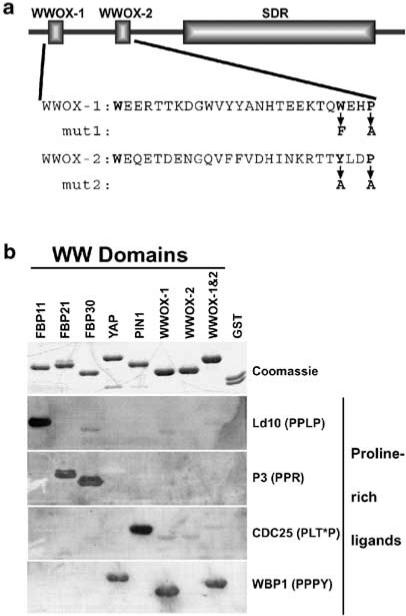
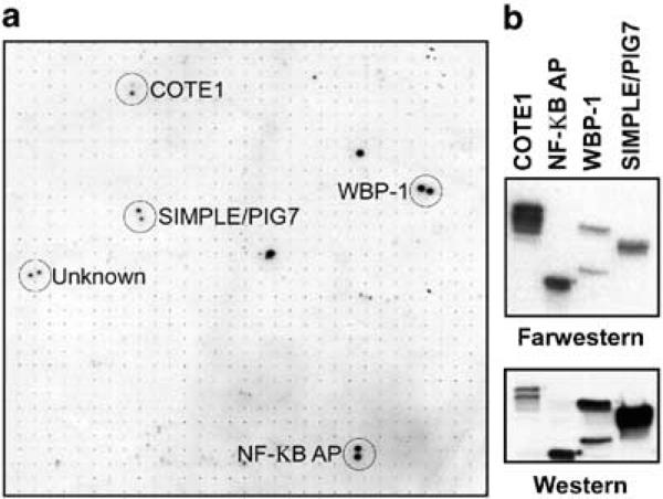
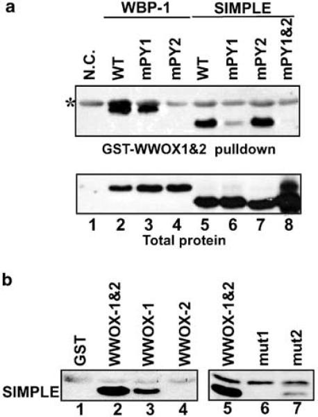
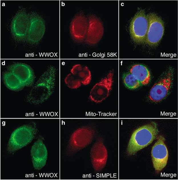
WWOX, the gene that maps to common chromosomal fragile site FRA16D, is frequently affected by aberrations in multiple types of cancers. WWOX encodes a 46 kDa protein that contains two WW domains and a short-chain oxidoreductase (SDR) domain. We recently demonstrated that ectopic expression of WWOX inhibits xenograft tumor growth of tumorigenic breast cancer cells. Little is known of the biochemical function(s) of WWOX. The SDR domain is predicted to be involved in sex-steroid metabolism and the WW domains are likely involved in protein-protein interactions. In this report, we identify the specific proline-rich ligand for WWOX as PPXY and show that the amino-terminal WW domain is responsible for this interaction. Using the WWOX WW domains as a probe, we screened high-density protein arrays and identified five candidate-binding partners. The binding to one of these candidates, small membrane protein of the lysosome/late endosome (SIMPLE), was further analysed, and we observed that a specific PPSY motif in the SIMPLE amino-acid sequence was required to interact with the amino-terminal WW domain of WWOX. In addition, immunofluorescence staining demonstrated that endogenous WWOX and SIMPLE co-localize to perinuclear compartments of MCF-7 human breast cancer cells. These studies demonstrate that WWOX contains a Group I WW domain that binds known cellular proteins containing the specific ligand PPXY. Identification and characterization of WWOX interacting proteins will lead to an understanding of the biological functions of WWOX in normal and tumor cells.


Figures

Figure 1
Figure 1
WWOX WW domain I binds the PPXY ligand. (a) Full-length WWOX protein is shown schematically, highlighting the WW domains and the SDR domain. Below the schematic are the amino-acid sequences of the WW domains. Mut1 and mut2 show the amino-acid changes made in GST–WWOX-1 and -2 to inactivate the WWOX-1 and WWOX-2 domains, respectively. (b) Purified GST-fusion proteins (top panel) containing the WW domains from several different proteins were probed with each of four proline-rich ligands (bottom four panels). Equal amounts of each GST-fusion protein and GST alone were separated by SDS–PAGE and stained with Coomassie blue to visualize protein bands (Coomassie) or transferred to PVDF membrane for ligand binding. The biotinylated peptide ligands (PPLP, PPR, PLT*P, and PPPY) were bound to streptavidin-conjugated HRP prior to incubation with the membrane-bound proteins. Ligands bound by GST-fusion proteins were visualized by ECL. The complete amino-acid sequences of the peptide ligands are given in Materials and methods. T* = phosphothreonine
Figure 2
Figure 2
Identification of candidate WWOX interacting proteins. (a) High-density protein arrays were probed with a 32P radiolabeled WW domain. Proteins bound to the WWOX WW domains were visualized by autoradiography and were identified by mapping their coordinates on the array grid. A partial view of the array is shown with duplicate positive colonies circled. The name of each protein identified by BLAST search is shown. (b) The interaction of the WWOX WW domains with the identified proteins was confirmed by far-Western analysis of bacterial expressed candidate proteins. Each candidate protein was bound to nickel agarose beads under denaturing conditions, separated by SDS–PAGE and transferred to PVDF membranes. Membranes were analysed by Western blotting (bottom panel) using antibodies that recognized the histidine tag, and by far-Western analysis (top panel) using radiolabeled WWOX WW domains as a probe
Figure 3
Figure 3
(a) WWOX interacts with specific PPXY motifs of candidate interacting proteins. Site-directed mutagenesis was used to change the tyrosine of PPXY to alanine of SIMPLE (lanes 5–8) and WBP-1 (lanes 2–4). Additionally, a double mutant of SIMPLE was created (lane 8). To determine protein interactions, total protein extracts from bacteria expressing the wild-type and mutant proteins were used for GST pull-down assays. Proteins pulled down by GST–WWOX-1 and -2 were separated by SDS–PAGE, transferred to PVDF membranes and visualized by Western blotting using antibody to the histidine tag (top panel). Equal amounts of expressed proteins were determined by Western blotting of total protein extracts (bottom panel). Total protein extracts from the parental bacterial strain that does not express any histidine-tagged proteins were used as the negative control (lane 1). *Band due to nonspecific binding of the antibody to the GST protein. (b) The first WW domain is required for interaction with endogenously expressed SIMPLE. Whole-cell extracts from the breast cancer cell line MCF-7 were used for GST pull-down assays using GST fused to each individual WWOX WW domain and to GST–WWOX-1 and -2 containing altered amino acids in each of the WW domains. Proteins pulled down by the indicated GST-fusion proteins were separated by SDS–PAGE, transferred to PVDF and analysed by Western blotting with antibody that specifically recognizes SIMPLE. Left panel: Binding to the complete WWOX WW domain (GST–WWOX-1 and -2, lane 2) and to the individual WWOX WW domains (GST–WWOX-1 and GST–WWOX-2, lanes 3–4, respectively). The observed binding by endogenous SIMPLE was specific for WWOX, as shown by the GST pull-down with GST alone (lane 1). Right panel: Binding to the complete WW domain (GST–WWOX-1 and -2, lane 5) and to the mutant WW domains (mut1 and mut2, lanes 6 and 7, respectively). Each WW domain had two amino acids changed in the context of GST–WWOX-1 and -2 (Figure 1)
Figure 4
Figure 4
WWOX and SIMPLE partially co-localize to the Golgi apparatus. Panels a–c: Endogeous WWOX co-localizes with the 58 kDa Golgi marker in MCF-7 cells. Note full overlap of the fluorescence signals (panel c, yellow color). Panels d–f: Immunofluorescence detection of WWOX and fluorescent staining of mitochondria. As can be observed, no overlap is evident in the merge image (panel f). Panels g–i: Endogenous WWOX partially co-localizes with SIMPLE. Immunofluorescence detection of WWOX (panels a, d and g) was carried out using affinity-purified anti-WWOX rabbit polyclonal antiserum and Alexafluor 488 conjugated anti-rabbit secondary antibody. Immunofluorescence detection of 58 kDa Golgi marker (panel b) was carried out using mouse monoclonal anti-58 kDa and Alexafluor 647-conjugated anti-mouse secondary antibody. Immunofluorescence detection of SIMPLE (panel h) was carried out using mouse monoclonal anti-SIMPLE antibody and Alexafluor 647 secondary antibody

Similar articles

Cited by

References

    1. Bedford MT, Reed R, Leder P. Proc. Natl. Acad. Sci. USA. 1998;95:10602–10607. - PMC - PubMed
    1. Bedford MT, Sarbassova D, Xu J, Leder P, Yaffe MB. J. Biol. Chem. 2000;275:10359–10369. - PubMed
    1. Bednarek AK, Keck-Waggoner CL, Daniel RL, Laflin KJ, Bergsagel PL, Kiguchi K, Brenner AJ, Aldaz CM. Cancer Res. 2001;61:8068–8073. - PubMed
    1. Bednarek AK, Laflin KJ, Daniel RL, Liao Q, Hawkins KA, Aldaz CM. Cancer Res. 2000;60:2140–2145. - PubMed
    1. Bussow K, Cahill D, Nietfeld W, Bancroft D, Scherzinger E, Lehrach H, Walter G. Nucleic Acids Res. 1998;26:5007–5008. - PMC - PubMed

Publication types