. 2004 Apr 13;43(14):4196-205.
doi: 10.1021/bi035992a.

DNA damage recognition of mutated forms of UvrB proteins in nucleotide excision repair

Affiliations

DNA damage recognition of mutated forms of UvrB proteins in nucleotide excision repair

Yue Zou et al. Biochemistry. .

Erratum in

  • Biochemistry. 2004 Jun 1;43(21):6832

Abstract

The DNA repair protein UvrB plays an indispensable role in the stepwise and sequential damage recognition of nucleotide excision repair in Escherichia coli. Our previous studies suggested that UvrB is responsible for the chemical damage recognition only upon a strand opening mediated by UvrA. Difficulties were encountered in studying the direct interaction of UvrB with adducts due to the presence of UvrA. We report herein that a single point mutation of Y95W in which a tyrosine is replaced by a tryptophan results in an UvrB mutant that is capable of efficiently binding to structure-specific DNA adducts even in the absence of UvrA. This mutant is fully functional in the UvrABC incisions. The dissociation constant for the mutant-DNA adduct interaction was less than 100 nM at physiological temperatures as determined by fluorescence spectroscopy. In contrast, similar substitutions at other residues in the beta-hairpin with tryptophan or phenylalanine do not confer UvrB such binding ability. Homology modeling of the structure of E. coli UvrB shows that the aromatic ring of residue Y95 and only Y95 directly points into the DNA binding cleft. We have also examined UvrB recognition of both "normal" bulky BPDE-DNA and protein-cross-linked DNA (DPC) adducts and the roles of aromatic residues of the beta-hairpin in the recognition of these lesions. A mutation of Y92W resulted in an obvious decrease in the efficiency of UvrABC incisions of normal adducts, while the incision of the DPC adduct is dramatically increased. Our results suggest that Y92 may function differently with these two types of adducts, while the Y95 residue plays an unique role in stabilizing the interaction of UvrB with DNA damage, most likely by a hydrophobic stacking.


Figures

F<sc>igure</sc> 1:
Figure 1:
Structures of DNA substrates used in the present study. The bold X in the sequences represents the single adducted nucleotide. The arrows indicate the major incision sites of UvrABC.
F<sc>igure</sc> 2:
Figure 2:
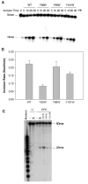
Incision of BPDE and DPC substrates by UvrABC nuclease with wild-type (WT) or various mutant UvrBs. Panel A: 5′-Terminally labeled 50 bp substrates (3 nM) containing a (+)-cis-BPDE adduct were incubated with UvrABC (UvrA, 15 nM, UvrB or mutant, 250 nM, and UvrC, 50 nM) in the UvrABC buffer at 37 °C for the indicated time periods. The incision products were analyzed on a 12% polyacrylamide sequencing gel. The 50mer represents the intact top strand DNA (labeled), while the 18mer represents the products of 5′-incision. Panel B: Rates of UvrABC incisions of the substrate with wild-type UvrB and UvrB mutants. The error bars represent standard deviations of the measurements. Panel C: Double strand labeled substrates containing a DPC adduct were incised under the same experimental conditions except that the incubation time was 60 min. The 60mer represents the intact bottom strand DNA which was also 5′-terminally labeled for the purpose of quantification, while the 23mer is the product of the 5′-incision. The U denotes the 60 bp DNA containing a uracil instead of DPC as a control.
F<sc>igure</sc> 3:
Figure 3:
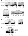
Direct binding of UvrB and mutant proteins to various DNA substrates. Panel A: UvrB or UvrB mutants (800 nM, or as indicated by +, 200 nM, and ++, 600 nM) were incubated with DNA substrates (3 nM) at 37 °C for 15 min in the UvrABC buffer without ATP. The binding products were analyzed on a 3.5% native polyacrylamide gel. The notation free DNA represents the unbound DNA substrates, and DNA-UvrB represents the complexes formed between DNA and the UvrB mutants. The F-30bp-bubble substrate stands for fluorescein-30 bp bubble. Panel B: Same as in panel A except that the symbols + and ++ represent UvrB concentrations of 100 and 400 nM, respectively. Panel C: Labeled BPDE-DNA 50 bp substrates were incubated with UvrA (15 nM) and UvrB/mutants (100 nM) at 37 °C for 15 min in UvrABC buffer with 1 mM ATP. The samples were analyzed on a 3.5% native polyacrylamide gel running with TBE buffer in the presence of 1 mM ATP and 10 mM MgCl2. F, CPD, and ND stand for fluorescein, cyclobutane pyrimidine dimer, and nondamaged DNA, respectively.
F<sc>igure</sc> 4:
Figure 4:
Emission spectra of mutant UvrB proteins. The tryptophan fluorescence of the proteins (1 μM) was recorded (in arbitrary units) at 25 °C with excitation at 295 nm in 200 uL of UvrABC buffer solution in the absence of ATP (50 mM Tris-HCl, pH 7.5, 50 mM KCl, 10 mM MgCl2, 5 mM DTT). The slit widths were set at 3 and 5 nm for excitation and emission beams, respectively.
F<sc>igure</sc> 5:
Figure 5:
Fluorescence titration of fluorescein-DNA substrate with UvrB(Y95W). Fluorescein-DNA substrate (0.1 μM) was titrated with UvrB(Y95W) at 37 °C in the UvrABC buffer without ATP by measuring the emission of fluorescein at 520 nm (excitation at 492 nm). The slit widths were set at 1 and 5 nm for excitation and emission beams, respectively.
F<sc>igure</sc> 6:
Figure 6:
Binding of UvrB and mutants to the DNA-protein cross-link adduct. UvrB or UvrB mutants of 400 (+) or 800 (++) nM were incubated with DPC substrate (3 nM) at 37 °C for 15 min in the UvrABC buffer without ATP. The binding products were analyzed on a 3.5% native polyacrylamide gel. Free DPC represents the unbound DPC-DNA substrate, and the DPC-UvrB represents the complex formation between DNA and mutants.
F<sc>igure</sc> 7:
Figure 7:
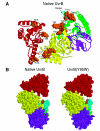
Modeled structures of E. coli UvrB and its mutant Y95W. Structures were generated by homology modeling based on the 2.6 Å crystal structure of B. caldotenax UvrB (see Experimental Procedures). The quality of the modeled structures was evaluated by the PROCHECK program, indicating that about 98% of the residues are in the most favored and the additionally allowed regions of the Ramachandran plot. Domains of the structures are colored in yellow for domain 1a, green for domain 1b, purple for domain 2, red for domain 3, and cyan for the β-hairpin. Panel A shows the structure of native UvrB (ribbon). Aromatic residues (space-filling model) at the cleft are colored in gold. Panel B presents the space-filling structures of native UvrB and the UvrB(Y95w) mutant for comparison mainly at residue 95. Y95 (native UvrB) and W95 (mutant) are colored in gold.

Similar articles

Cited by

References

    1. Van Houten B. Microbiol. Rev. 1990;54:18–51. - PMC - PubMed
    1. Sancar A. Annu. Rev. Biochem. 1996;65:43–81. - PubMed
    1. Wood RD. Biochimie. 1999;81:39–44. - PubMed
    1. Minko IG, Zou Y, Lloyd RS. Proc. Natl. Acad. Sci. U.S.A. 2002;99:1905–1909. - PMC - PubMed
    1. Zou Y, Luo C, Geacintov NE. Biochemistry. 2001;40:2923–2931. - PubMed

Publication types

MeSH terms