. 2004 Apr;52(2):573-87.
doi: 10.1111/j.1365-2958.2004.04000.x.

Genetic evidence that the Vibrio cholerae monolayer is a distinct stage in biofilm development

Affiliations

Genetic evidence that the Vibrio cholerae monolayer is a distinct stage in biofilm development

Sudha Moorthy et al. Mol Microbiol. 2004 Apr.

Abstract

Biofilm development is conceived as a developmental process in which free swimming cells attach to a surface, first transiently and then permanently, as a single layer. This monolayer of immobilized cells gives rise to larger cell clusters that eventually develop into the biofilm, a three-dimensional structure consisting of large pillars of bacteria interspersed with water channels. Previous studies have shown that efficient development of the Vibrio cholerae biofilm requires a combination of pili, flagella and exopolysaccharide. Little is known, however, regarding the requirements for monolayer formation by wild-type V. cholerae. In this work, we have isolated the wild-type V. cholerae monolayer and demonstrated that the environmental signals, bacterial structures, and transcription profiles that induce and stabilize the monolayer state are unique. Cells in a monolayer are specialized to maintain their attachment to a surface. The surface itself activates mannose-sensitive haemagglutinin type IV pilus (MSHA)-mediated attachment, which is accompanied by repression of flagellar gene transcription. In contrast, cells in a biofilm are specialized to maintain intercellular contacts. Progression to this stage occurs when exopolysaccharide synthesis is induced by environmental monosaccharides. We propose a model for biofilm development in natural environments in which cells form a stable monolayer on a surface. As biotic surfaces are degraded with subsequent release of carbohydrates, the monolayer develops into a biofilm.


Figures

Fig. 1
Fig. 1
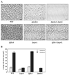
Monolayer formation by wild-type V. cholerae (WT, MO10) and ΔvpsA (PW396), ΔflaA (PW412), and Δ mshA (PW361) mutants after incubation in MM alone at 27°C for 24 h. A. Phase-contrast micrographs of monolayers. Bar = 10 µm. B. Total surface area covered by surface-attached cells. Observations represent the mean of three separate experiments.
Fig. 2
Fig. 2
Effect of α-methyl mannoside (AMM) on surface association by wild-type V. cholerae (WT, MO10) and a ΔflaA mutant (PW412) in MM alone. A. Phase-contrast micrographs of monolayers before (MM) and after (MM + AMM) treatment with AMM. Bar = 10 µm. B. Per cent total surface area covered by monolayer cells before and after treatment with AMM as noted in the key. Observations represent the mean of three separate experiments.
Fig. 3
Fig. 3
Biofilm formation by wild-type V. cholerae (WT, MO10) and ΔvpsA (PW396), ΔflaA (PW412), ΔmshA (PW361), ΔmshAΔvpsL (PW448), and ΔflaAΔvpsL (PW450) mutants in MM supplemented with mannose. A. Phase-contrast micrographs of biofilm formation. Bar = 10 µm. B. Comparison of mean cluster size of surface-associated cells in MM alone and supplemented with mannose as noted in the key. Observations represent the mean of three separate experiments.
Fig. 4
Fig. 4
Effect of α-methyl mannoside (AMM) on surface association by wild-type V. cholerae (WT, MO10) and a ΔflaA mutant (PW412) in MM supplemented with mannose. A. Phase-contrast micrographs before (MM + M) and after (MM + M + AMM) treatment with AMM. Bar = 10 µm. B. Percentage of the total surface area covered by biofilm-associated cells before and after treatment with AMM as noted in the key. Observations represent the mean of three separate experiments.
Fig. 5
Fig. 5
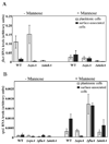
Normalized levels of mRNA transcripts in planktonic (grey bars) or surface-associated (black bars) wild-type V. cholerae (WT, MO10) and ΔvpsA (PW396), ΔflaA (PW412) and ΔmshA (PW361) mutant cells. Cells were incubated in MM alone (−Mannose) or supplemented with mannose (+Mannose). A. Quantification of flaA mRNA. B. Quantification of vpsL mRNA. Observations represent the mean of at least three separate experiments.
Fig. 6
Fig. 6
A model for V. cholerae biofilm development in natural environments. Planktonic V. cholerae enter the monolayer stage upon encounter with a surface through the action of MSHA. This is accompanied by the loss of the flagellum. Degradation and transport of oligosaccharides and monosaccharides from the surface provide the signal for progression to the biofilm stage.

References

    1. Ali A, Rashid MH, Karaolis DK. High-frequency rugose exopolysaccharide production by Vibrio cholerae. Appl Environ Microbiol. 2002;68:5773–5778. - PMC - PubMed
    1. Allison DG, Ruiz B, SanJose C, Jaspe A, Gilbert P. Extracellular products as mediators of the formation and detachment of Pseudomonas fluorescens bio-films. FEMS Microbiol Lett. 1998;167:179–184. - PubMed
    1. Bechet M, Blondeau R. Factors associated with the adherence and biofilm formation by Aeromonas caviae on glass surfaces. J Appl Microbiol. 2003;94:1072–1078. - PubMed
    1. Bomchil N, Watnick P, Kolter R. Identification and characterization of a Vibrio cholerae gene, mbaA, involved in maintenance of biofilm architecture. J Bacteriol. 2003;185:1384–1390. - PMC - PubMed
    1. Bonet R, Simon-Pujol MD, Congregado F. Effects of nutrients on exopolysaccharide production and surface properties of Aeromonas salmonicida. Appl Environ Microbiol. 1993;59:2437–2441. - PMC - PubMed

Publication types

MeSH terms