. 2004 Apr 5:5:13.
doi: 10.1186/1471-2121-5-13.

Effects of "second-hand" smoke on structure and function of fibroblasts, cells that are critical for tissue repair and remodeling

Affiliations

Effects of "second-hand" smoke on structure and function of fibroblasts, cells that are critical for tissue repair and remodeling

Lina S Wong et al. BMC Cell Biol. .

Abstract

Background: It is known that "second-hand" cigarette smoke leads to abnormal tissue repair and remodelling but the cellular mechanisms involved in these adverse effects are not well understood. Fibroblasts play a major role in repair and remodelling. They orchestrate these processes by proliferating, migrating, and secreting proteins such as, cytokines, growth factors and extracellular matrix molecules. Therefore, we focus our studies on the effects of "second-hand" cigarette smoke on the structure and function of these cells.

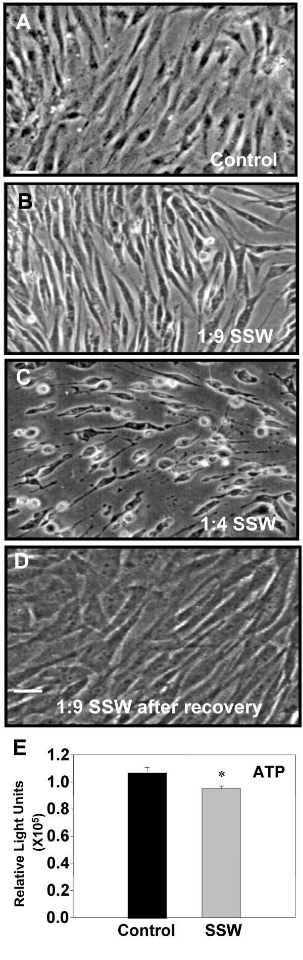
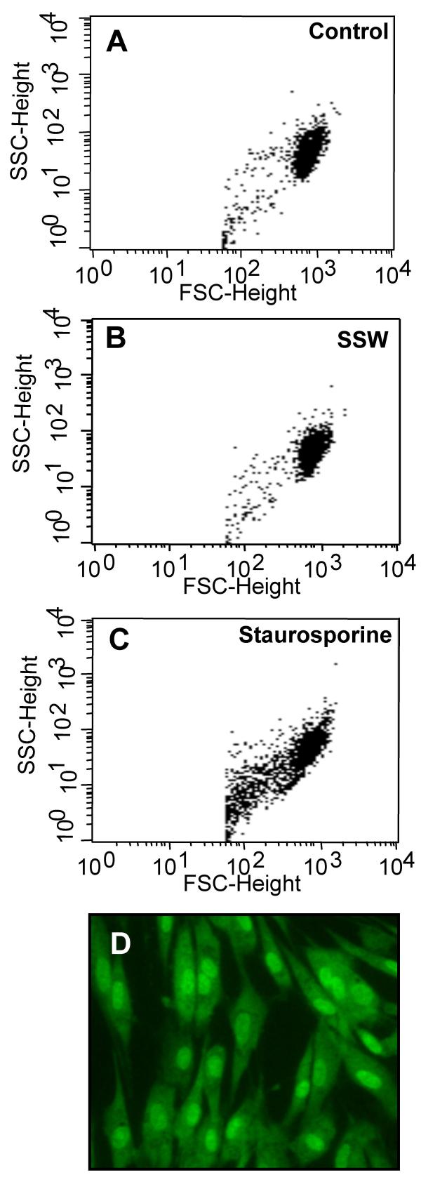
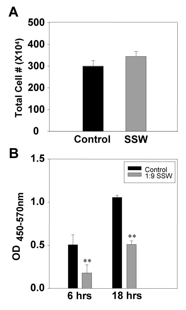
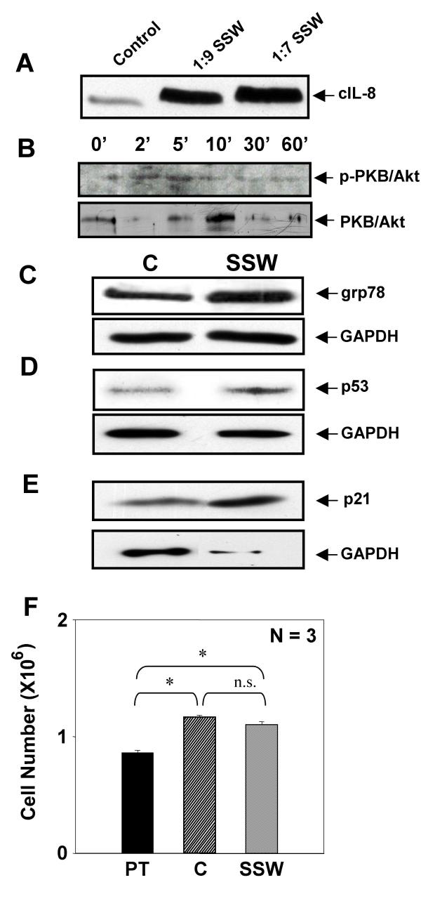
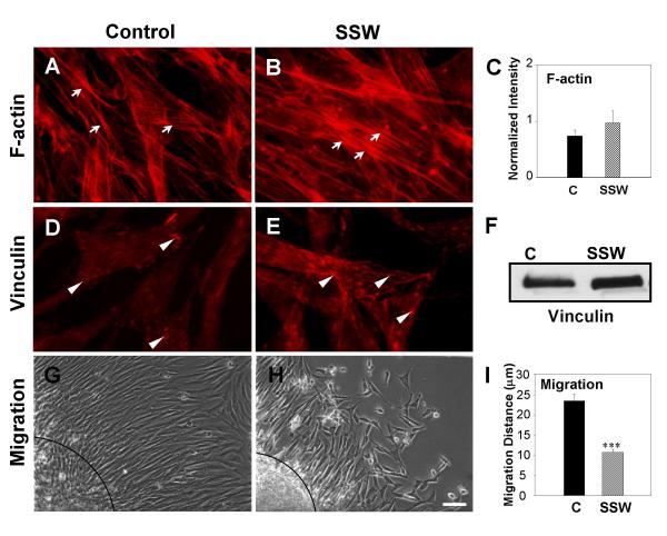
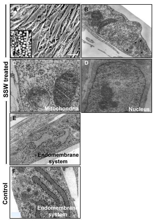
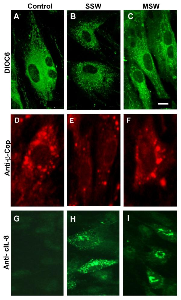
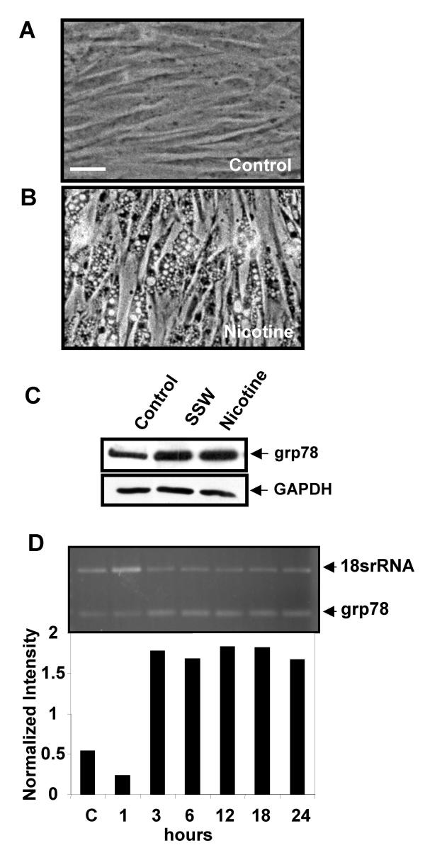
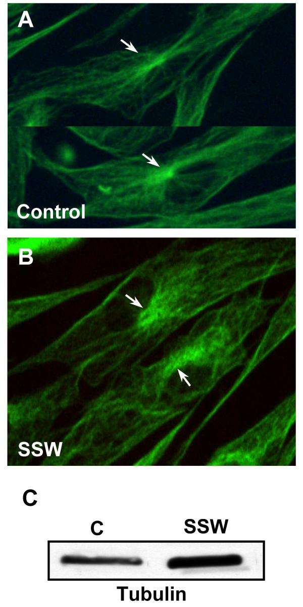
Results: We used sidestream whole (SSW) smoke, a major component of "second-hand" smoke, primary embryonic fibroblasts, cells that behave very much like wound fibroblasts, and a variety of cellular and molecular approaches. We show that doses of smoke similar to those found in tissues cause cytoskeletal changes in the fibroblasts that may lead to a decrease in cell migration. In addition, we also show that these levels of cigarette smoke stimulate an increase in cell survival that is reflected in an increase and/or activation of stress/survival proteins such as cIL-8, grp78, PKB/Akt, p53, and p21. We further show that SSW affects the endomembrane system and that this effect is also accomplished by nicotine alone.

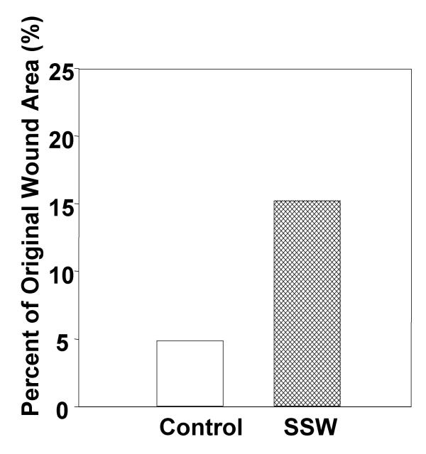
Conclusions: Taken together, our results suggest that: (i) SSW may delay wound repair because of the inability of the fibroblasts to migrate into the wounded area, leading to an accumulation of these cells at the edge of the wound, thus preventing the formation of the healing tissue; (ii) the increase in cell survival coupled to the decrease in cell migration can lead to a build-up of connective tissue, thereby causing fibrosis and excess scarring.


Figures

Figure 1
Figure 1
Phase-contrast microscopy analysis of sidestream whole (SSW) smoke treated primary fibroblasts. Cells were treated with different doses of SSW for 18 hours. Control cells were kept in serum-free medium for the same time period because the smoke solutions are diluted in serum-free medium. (A) Untreated cells were spread out, confluent, contact inhibited, and showed prominent nuclei. (B) Cells treated with 1:9 (SSW:media) smoke dilution became elongated and separated from each other. (C) Cells treated with 1:4 SSW smoke rounded up and showed signs of cell death. (D) Cells treated with 1:9 SSW smoke solution recovered quickly; within a few hours of being in complete media they were back to normal morphology. (E) ATP assays: cells were plated in a 96 well ELISA plate, allowed to reach confluency, and treated for 18 hours with SSW. Smoke-treated cells showed a decrease in ATP production, but the overall ATP level remained high. Pictures are representative of at least 3 experiments performed with different batches of primary cells. Scale bar = 20 μm.
Figure 2
Figure 2
Flow cytometric analysis of cells treated with SSW smoke. Cells were treated with 1:9 SSW smoke or with staurosporine and analyzed by flow cytometry to determine the forward- and side-scattering properties of the cells. (A&B) Untreated and SSW-treated cells show similar pattern of forward- and side-scattering properties. (C) Staurosporine treated cells (positive control) showed more cells with lower forward-scattering properties than either control or SSW-treated cells suggesting that SSW smoke is not causing cell death and that the overall structure of the cell is normal. The graphs represent 10,000 events. (D) Acridine-orange and ethidium bromide staining; cells showed normal morphology, and no blebbing of the nucleus or plasma membrane was observed. SSC = side-scatter, FSC = forward-scatter. Figures are representative of at least 3 repeated studies. Scale bar = 20 μm.
Figure 3
Figure 3
Effects of SSW smoke on fibroblast growth. (A) Cells were treated for 18 hours with SSW smoke solution and the total cell number was counted using a Coulter counter with a specified particle size of 7 μm to 20 μm. There was no significant difference between controls and treated cells. (B) Primary fibroblasts were plated in 96-well plates and allowed to grow to confluency and BrdU alone or BrdU plus SSW were added to the cultures and the cells were allowed to incorporate the BrdU for the indicated time points. At both 6 and 18 hours, SSW-treated cells showed a significant decrease in BrdU incorporation when compared to the control. Experiments are performed at least two times with different batches of primary fibroblasts. OD = Optical Density.
Figure 4
Figure 4
SSW smoke stimulates stress response proteins and cell survival. SSW stimulated an increase in immediate early stress-response proteins as assayed by immunoblotting analysis. (A) cIL-8 was stimulated in a dose-dependent manner. (B) PKB/Akt was phosphorylated/activated by 5 minutes. (C-E) The levels of grp78 (C), p53 (D), and p21 (E) were all increased upon stimulation with SSW smoke. GAPDH was used as a marker for levels of sample loading. Because the supernatant does not contain GAPDH, we verified equal loading for cIL-8 by using coomassie blue staining of identical samples. (F) Cells were treated for 18 hours, allowed to recover in fresh medium for 24 hours, treated again for 18 hours and then counted using a Coulter counter. The number of cells after SSW treatment was comparable to that of the control, suggesting that cells survived well even though they were cultured in the presence of SSW smoke. For immunoblotting analysis, cells were treated and lysates separated using SDS-PAGE. Each figure is a representative of at least 2 experiments. PT = Pretreatment; N = 3 indicates 3 samples per experimental group.
Figure 5
Figure 5
Effects of SSW smoke on microfilaments and focal adhesion plaques. Cells were treated with 1:9 smoke dilutions and different markers were analyzed. (A, B) Rhodamine-phalloidin labeling of F-actin showed that treated fibroblasts have more F-actin staining and the stress fibers appeared thicker. (C) The increase in F-actin was confirmed by staining the cells and measuring the amount of rhodamine-phalloidin present in the cells using a fluorimeter at 550–580 nm. (D, E) Fluorescence images of cells treated with SSW smoke and labeled for the focal adhesion plaque protein, vinculin. Smoke treated cells showed an increased in focal adhesion plaque formation compare with control cells. (F) Immunoblot analysis for vinculin confirms that SSW stimulates an increase in vinculin levels. For equal loading of protein in the immunoblots, please refer to grp78 blots in Fig. 4C; the same membrane was used to reprobe for the protein shown in this figure. (G, H) Effects of SSW on cell migration. Cells were plated inside cloning rings and allowed to adhere for 3 hours to form a "ring of cells". After marking the edge of the ring, the cells were treated and allowed to migrate for 24 hours and the migrated distance was measured from the edge of the ring to the migrating front of the cells. The treated cells showed a decrease in cell migration. (I) Quantification of the extent of inhibition of cell migration by SSW smoke. Data are representative of six different points along the circle. Dashed lines in G&H demark the edge of the "circle of cells". All experiments were performed at least 3 times with different batches of primary cells. Scale bar = 20 μm.
Figure 6
Figure 6
Microscopic analysis of cells treated with SSW smoke. (A) SSW-treated cells developed numerous vacuoles in the cytosol. Inset shows a higher magnification of the vacuoles. (B-F) Cells treated with SSW for 4 hours were fixed and prepared for Transmission Electron Microscopy (TEM). We observed that the cells are still morphologically intact (B) except for the endomembrane system, which is beginning to show swelling (arrows). However, most organelles, such as the mitochondria (C) and the nucleus (D) look normal, whereas the endomembrane system is irregularly shaped and shows signs of swelling (E). (F) Endomembrane system of untreated cells. Scale bars = 20 μm in (A), 5 μm in (B) and 0.6 μm in (C-F).
Figure 7
Figure 7
SSW smoke affects the endomembrane network. Cells were exposed to 4 or 8 hours of smoke treatment (SSW or MSW) and prepared for fluorescence imaging. (A) Confocal microscopy analysis of DIOC6 stained cells. Untreated cells show normal ER network concentrated around the nucleus and also spread out to the periphery of the cell. (B) SSW treated cells show that the ER is fragmented when compared with the control and is only found around the nucleus. (C) MSW-treated cells show similar ER morphology to that of the control. (D-F) To ascertain the status of the Golgi network, we used anti-β-COP monoclonal antibody for immunocytochemistry, which specifically labels the Golgi network. (D) In untreated cells, the staining of the Golgi surrounds the nucleus and vesicles are seen all over the cytosol. (E) In SSW-treated cultures, the cells have many fewer Golgi vesicle staining. (F) MSW treated cells show an organization similar to that found in the control. (G-I) Fibroblasts were treated as above and immunolabeled for cIL-8. (G) In untreated cells, the expression of cIL-8 is barely present because this protein is only expressed when it is stress-induced. (H) SSW-treated cells were observed to have no perinuclear staining; instead, the endomembrane system was dispersed throughout the cytosol. (I) MSW-treated cells show that the cIL-8 is being produced in the endomembrane system near the nucleus where this normally occurs. Pictures are representative of 2 different experiments. Scale bars = 50 μm in (A-F) and 30 μm in (G-I).
Figure 8
Figure 8
Nicotine mimics the effects of SSW smoke on the endomembrane. (A&B) Phase contrast microscopy of cells treated with 1.5 mM nicotine for 4 hours showed vacuolation similar to that found in fibroblasts treated with SSW smoke (Compare with Fig. 6A). (C) Western blot analysis showing that nicotine stimulated grp78 expression, an ER specific stress response protein, to similar levels as SSW. GAPDH shows equal loading of cell lysate. (D) RT-PCR of nicotine-treated cells showed that this component of smoke stimulated grp78 expression. Data are representative of at least 3 different studies with 3 different batches of primary cells. Scale bar = 20 μm.
Figure 9
Figure 9
The effects of SSW on microtubules. (A, B) Fluorescence images of microtubules labeled with an antibody to tubulin. (A) Control cells showed a characteristic, brightly labeled microtubule organizing center (MTOC=centrosome) with microtubules radiating outward throughout the cytoplasm. (B) SSW-treated cells showed a much less orderly extension of the tubules, and the MTOC were much less organized than in the control cells. (C) Immunoblot analysis showed an increase in tubulin after smoke treatment. For equal loading of protein in the immunoblots, please refer to grp78 blots in Fig. 4C; the same membrane was used to reprobe for the protein shown in this figure.
Figure 10
Figure 10
Effect of SSW smoke on wound closure. Mice were wounded with a 5 μm biopsy punch, pictures were taken at 5 and 7 days with 7.5× magnification. The areas of the wounds were quantified using Scion Image Software. By 7 days, the wounds of the control animals were approximately 95% closed whereas SSW wounds were only 85% closed. Data show the results of two representative experiments.

References

    1. Pirkle JL, Flegal KM, Bernert JT, Brody DJ, Etzel RA, Maurer KR. Exposure of the US population to environmental tobacco smoke: the Third National Health and Nutrition Examination Survey, 1988 to 1991. Jama. 1996;275:1233–1240. doi: 10.1001/jama.275.16.1233. - DOI - PubMed
    1. EPA Respiratory Health Effects of Passive Smoking: Lung Cancer and Other Disorders. 1992. pp. 3.1–3.1.
    1. Lofroth G. Environmental tobacco smoke: overview of chemical composition and genotoxic components. Mutat Res. 1989;222:73–80. - PubMed
    1. Frick WG, Seals R. R., Jr. Smoking and wound healing: a review. Tex Dent J. 1994;111:21–23. - PubMed
    1. Silverstein P. Smoking and wound healing. Am J Med. 1992;93:22S–24S. - PubMed

Publication types

Substances