Comparative Study
. 2004 Apr;70(4):2349-53.
doi: 10.1128/AEM.70.4.2349-2353.2004.

Subtilosin production by two Bacillus subtilis subspecies and variance of the sbo-alb cluster

Affiliations
Comparative Study

Subtilosin production by two Bacillus subtilis subspecies and variance of the sbo-alb cluster

Torsten Stein et al. Appl Environ Microbiol. 2004 Apr.

Abstract

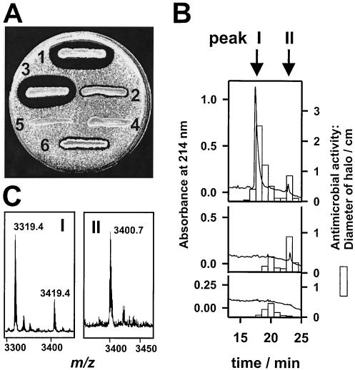
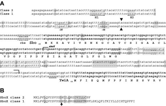
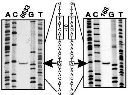
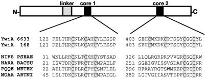
Eight different Bacillus subtilis strains and Bacillus atrophaeus were found to produce the bacteriocin subtilosin A. On the basis of the subtilosin gene (sbo) sequences two distinct classes of B. subtilis strains were distinguished, and they fell into the two B. subtilis subspecies (B. subtilis subsp. subtilis and B. subtilis subsp. spizizenii). The entire sequence of the subtilosin gene cluster of a B. subtilis subsp. spizizenii strain, B. subtilis ATCC 6633, was determined. This sequence exhibited a high level of homology to the sequence of the sbo-alb gene locus of B. subtilis 168. By using primer extension analysis the transcriptional start sites of sbo in B. subtilis strains ATCC 6633 and 168 were found to be 47 and 45 bp upstream of the sbo start codon, respectively. Our results provide insight into the incipient evolutionary divergence of the two B. subtilis subspecies.


Figures

FIG. 1.
FIG. 1.
Analysis of B. subtilis peptide antibiotics. (A) Micrococcus luteus growth inhibition assay. Streak 1, B. subtilis wild-type strain ATCC 6633; streak 2, strain ATCC 6633 ΔspaS deletion mutant; streak 3, strain ATCC 6633 Δsbo deletion mutant; streak 4, strain ATCC 6633 ΔspaS/Δsbo deletion mutant; streak 5, strain 168 Δsbo deletion mutant; streak 6, B. subtilis wild-type strain 168. (B) Reversed-phase HPLC separation of B. subtilis ATCC 6633 culture supernatants. The flow rate was 0.4 ml/min, and 400-μl fractions were collected. Aliquots (20 μl) of the wild type (top panel), the ΔspaS mutant (middle panel), and the ΔspaS/Δsbo double mutant (bottom panel) were used to inhibit the growth of M. luteus. (C) Detail of MALDI-TOF mass spectra of peak I (m/z 3319.4 and 3419.4 correspond to subtilin and succinylated subtilin, respectively) and peak II (m/z 3400.7 corresponds to subtilosin A). The variance of the m/z measurements was ±0.2 Da.
FIG. 2.
FIG. 2.
Comparison of sbo alleles of two B. subtilis classes. (A) Subtilosin A-encoding gene sequence sbo and flanking regions and the derived amino acid sequence. rbs, standard prokaryotic ribosome binding site. Transcriptional start sites (see Fig. 3) are indicated by arrows, and the positions of derived −10 and −35 regions are indicated. H1 and H2 indicate a putative sigma factor H region located 80 to 100 bp upstream of the transcriptional start site of sbo. A putative termination loop is enclosed in a box. The putative open reading frame sboX (150 nucleotides) is indicated by brackets. (B) Alignment of the amino acid sequences of the putative SboX gene product. The arrow indicates the putative processing site (double Gly motif). Differences between the two alleles are indicated by shading.
FIG. 3.
FIG. 3.
Primer extension analysis of sbo: mapping of the transcriptional start site of sbo by primer extension analysis with RNA from B. subtilis ATCC 6633 and 168 (middle lanes). The outside lanes show the results of autoradiography of dideoxynucleotide sequencing reactions with primer AS26 complementary to the 5′ region of sbo. The transcriptional start sites are indicated by arrows, and the −10 regions are enclosed by boxes in the derived nucleotide sequence.
FIG. 4.
FIG. 4.
Domain structure of YwiA (AlbA): schematic representation of the putative domain structure of YwiA resulting from amino acid alignment of the sequences of B. subtilis ATCC 6633 and 168. Highly conserved regions (cores) are indicated by solid boxes; a gray box indicates a less conserved region. The first cysteine cluster (core 1) is highly homologous to active sites of proteins belonging to the MoaA-NifB-PqqE family carrying Fe-S centers, like NifB from Pseudomonas aeruginosa (NIFB PSEAE), NarA from B. subtilis (NARA BACSU), PqqE from Methylobacterium extorquens (PQQE METEX), and MoaA from Arthrobacter nicotinovorans (MOAA ARTNI).

References

    1. Ahimou, F., P. Jacques, and M. Deleu. 2000. Surfactin and iturin A effects on Bacillus subtilis surface hydrophobicity. Enzyme Microb. Technol. 27:749-754. - PubMed
    1. Anagnostopoulos, C., and J. Spizizen. 1961. Requirements for transformation in Bacillus subtilis. J. Bacteriol. 81:741-746. - PMC - PubMed
    1. Babasaki, K., T. Takao, Y. Shimonishi, and K. Kurahashi. 1985. Subtilosin A, a new antibiotic peptide produced by Bacillus subtilis 168: isolation, structural analysis, and biogenesis. J. Biochem. (Tokyo) 98:585-603. - PubMed
    1. Banerjee, S., and J. N. Hansen. 1988. Structure and expression of a gene encoding the precursor of subtilin, a small protein antibiotic. J. Biol. Chem. 263:9508-9514. - PubMed
    1. Birnboim, H. C., and J. Doly. 1979. A rapid alkaline extraction procedure for screening recombinant plasmid DNA. Nucleic Acids Res. 7:1513-1523. - PMC - PubMed

Publication types

Associated data