. 2004 May;142(1):192-202.
doi: 10.1038/sj.bjp.0705691. Epub 2004 Apr 5.

Functional evidence of a role for two-pore domain potassium channels in rat mesenteric and pulmonary arteries

Affiliations

Functional evidence of a role for two-pore domain potassium channels in rat mesenteric and pulmonary arteries

M J Gardener et al. Br J Pharmacol. 2004 May.

Abstract

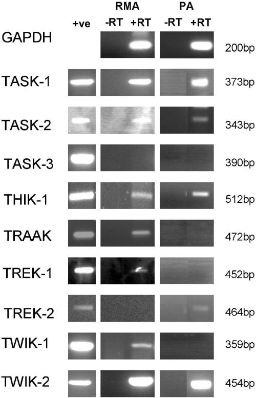
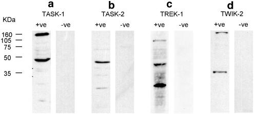
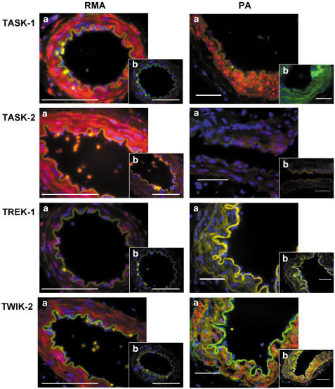
1. Experiments were performed to elucidate the mechanism by which alterations of extracellular pH (pH(o)) change membrane potential (E(M)) in rat mesenteric and pulmonary arteries. 2. Changing pH(o) from 7.4 to 6.4 or 8.4 produced a depolarisation or hyperpolarisation, respectively, in mesenteric and pulmonary arteries. Anandamide (10 microm) or bupivacaine (100 microm) reversed the hyperpolarisation associated with alkaline pH(o), shifting the E(M) of both vessels to levels comparable to that at pH 6.4. In pulmonary arteries, clofilium (100 microm) caused a significant reversal of hyperpolarisation seen at pH 8.4 but was without effect at pH 7.4. 3. K(+) channel blockade by 4-aminopyridine (4-AP) (5 mm), tetraethylammonium (TEA) (10 mm), Ba(2+) (30 microm) and glibenclamide (10 microm) depolarised the pulmonary artery. However, shifts in E(M) with changes in pH(o) remained and were sensitive to anandamide (10 microm), bupivacaine (100 microm) or Zn(2+) (200 microm). 4. Anandamide (0.3-60 microm) or bupivacaine (0.3-300 microm) caused a concentration-dependent increase in basal tone in pulmonary arteries. 5. RT-PCR demonstrated the expression of TASK-1, TASK-2, THIK-1, TRAAK, TREK-1, TWIK-1 and TWIK-2 in mesenteric arteries and TASK-1, TASK-2, THIK-1, TREK-2 and TWIK-2 in pulmonary arteries. TASK-1, TASK-2, TREK-1 and TWIK-2 protein was demonstrated in both arteries by immunostaining. 6. These experiments provide evidence for the presence of two-pore domain K(+) channels in rat mesenteric and pulmonary arteries. Collectively, they strongly suggest that modulation of TASK-1 channels is most likely to have mediated the pH-induced changes in membrane potential observed in these vessels, and that blockade of these channels by anandamide or bupivacaine generates a small increase in pulmonary artery tone.


Figures

Figure 1
Figure 1
Effects of changes in extracellular pH on membrane potential in mesenteric (a) and pulmonary arteries (b). Vessels were initially bathed in Tyrode's solution of pH 7.4. Reduction of extracellular pH (to pH 6.4) caused a marked depolarisation, while addition of alkaline Tyrode (pH 8.4) caused hyperpolarisation. Application of anandamide (10 μM) at either pH 8.4 or 7.4 returned EM to that seen with acidic pH0 (pH 6.4). (a1 and b1) Representative traces from mesentric (a1) and pulmonary (b1) arteries. (a2 and b2) Mean data ±s.e.m. derived from separate experiments (◊; n=3 or 4) on mesenteric and pulmonary arteries, respectively.
Figure 2
Figure 2
Effect of bupivacaine on changes in membrane potential with extracellular pH in mesenteric (a) and pulmonary arteries (b). Levcromakalim (LEV) was used as a positive control. Vessels were initially bathed in Tyrode's solution of pH 7.4. Reduction of extracellular pH (to pH 6.4) caused a marked depolarisation, while addition of alkaline Tyrode (pH 8.4) caused hyperpolarisation. Application of bupivacaine (100 μM) at pH 8.4 returned membrane potential to that seen in the presence of acidic Tyrode (pH 6.4), but was less effective at pH 7.4. (a1 and b1) Representative traces from mesenteric (a1) and pulmonary (b1) arteries are shown. (a2 and b2) Mean data±s.e.m. derived from separate experiments (◊; n=4) in mesenteric and pulmonary arteries are shown.
Figure 3
Figure 3
(a) Representative trace showing the effects of changes in extracellular pH on membrane potential in de-endothelialised pulmonary arteries. Vessels were initially bathed in Tyrode's solution of pH 7.4. Successful endothelial removal was assessed by application of 10 μM ACh. Reducing extracellular pH (to pH 6.4) caused marked depolarisation, while addition of alkaline Tyrode (pH 8.4) caused hyperpolarisation. This hyperpolarisation could be partially and reversibly inhibited by application of clofilium (100 μM). No effect could be seen upon application of clofilium (100 μM) when arteries where exposed to a Tyrode's solution of pH 7.4. (b) Mean±s.e.m. changes in membrane potential induced by alterations in pH0 and clofilium (100 μM) in four separate (◊) de-endothelialised pulmonary arteries.
Figure 4
Figure 4
Effects of changes in extracellular pH on membrane potential in pulmonary arteries. Vessels were initially bathed in Tyrode's solution of pH 7.4 prior to the addition of K+ channel inhibitor cocktail (CT) comprising 10 mM TEA, 10 μM glibenclamide, 5 mM 4-AP and 30 μM Ba2+. Cocktail application caused marked depolarisation. Reduction of pH (to pH 6.4) in the presence of cocktail produced a further depolarisation, while addition of alkaline Tyrode (pH 8.4) caused hyperpolarisation. While maintaining vessels in alkaline Tyrode in the presence of the cocktail, application of anandamide (10 μM) or Zn2+ returned membrane potential to values seen with acidic pH0 (pH 6.4). Representative traces from pulmonary arteries (a). Mean data±s.e.m. derived from four separate experiments (◊) on pulmonary arteries (b).
Figure 5
Figure 5
Effect of cumulative drug addition on resting tension elicited by anandamide and bupivacaine in small pulmonary arteries. Tension is presented as a mean percentage of that induced by 75 mM KCI PSS±s.e.m. (n=5–6).
Figure 6
Figure 6
Agarose gel electrophoresis of RT–PCR amplified with primers specific for GAPDH, TASK-1, TASK-2, TASK-3, THIK-1, TRAAK, TREK-1, TREK-2, TWIK-1 and TWIK-2. cDNA derived from rat brain or kidney was used as a positive control (+ve). Rat mesenteric and pulmonary artery mRNA was DNase treated and reverse transcribed. −RT controls were performed on DNase-treated mRNA that was not reverse transcribed. Products were sequenced to confirm identity.
Figure 7
Figure 7
Western blots of rat brain protein samples probed with anti -TASK-1, -TASK-2, -TREK-1 and -TWIK-2. Blots were performed with the primary antibody (+ve) or primary antibody that had been incubated with the antigenic peptide (−ve) prior to use. Theoretical weights of TASK-1, TASK-2, TREK-1 and TWIK-2 are 45, 55, 45 and 35 kDa, respectively.
Figure 8
Figure 8
Sections of rat mesenteric (RMA) and pulmonary artery (PA) stained with anti-TASK-1, -TASK-2, -TREK-1 and -TWIK-2. (a) Sections were stained with primary antibody and subsequently visualised using a Texas Red–conjugated secondary antibody. (b) Sections were labelled using the primary antibody that had been preincubated with the antigenic peptide. Nuclei were labelled blue with DAPI. Scale bars represent 100 μm.

References

    1. ARRIGHI I., LESAGE F., SCIMECA J.C., CARLE G.F., BARHANIN J. Structure, chromosome localization, and tissue distribution of the mouse twik K+ channel gene. FEBS Lett. 1998;425:310–316. - PubMed
    1. BRADFORD M.M. A rapid and sensitive method for the quantitation of microgram quantities of protein utilizing the principle of protein-dye binding. Anal. Biochem. 1976;72:248–254. - PubMed
    1. CHEN G., SUZUKI H., WESTON A.H. Acetylcholine releases endothelium-derived hyperpolarizing factor and EDRF from rat blood vessels. Br. J. Pharmacol. 1988;95:1165–1174. - PMC - PubMed
    1. CLAPP L.H., GURNEY A.M. Modulation of calcium movements by nitroprusside in isolated vascular smooth muscle cells. Pflugers Arch. 1991;418:462–470. - PubMed
    1. DECHER N., MAIER M., DITTRICH W., GASSENHUBER J., BRUGGEMANN A., BUSCH A.E., STEINMEYER K. Characterization of TASK-4, a novel member of the pH-sensitive, two-pore domain potassium channel family. FEBS Lett. 2001;492:84–89. - PubMed

Publication types

MeSH terms

LinkOut - more resources