. 2004 Apr 27;109(16):2023-9.
doi: 10.1161/01.CIR.0000127034.50006.C0. Epub 2004 Apr 5.

In vivo molecular imaging of acute and subacute thrombosis using a fibrin-binding magnetic resonance imaging contrast agent

Affiliations

In vivo molecular imaging of acute and subacute thrombosis using a fibrin-binding magnetic resonance imaging contrast agent

René M Botnar et al. Circulation. .

Abstract

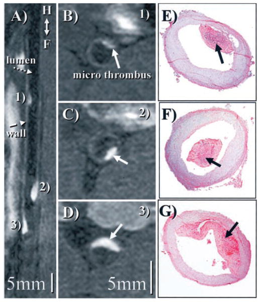
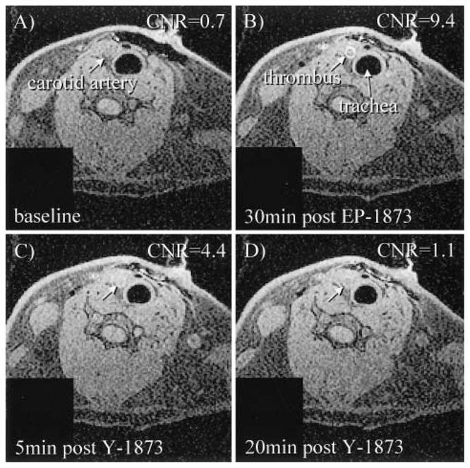
Background: Plaque rupture with subsequent thrombosis is recognized as the underlying pathophysiology of most acute coronary syndromes and stroke. Thus, direct thrombus visualization may be beneficial for both diagnosis and guidance of therapy. We sought to test the feasibility of direct imaging of acute and subacute thrombosis using MRI together with a novel fibrin-binding gadolinium-labeled peptide, EP-1873, in an experimental animal model of plaque rupture and thrombosis.

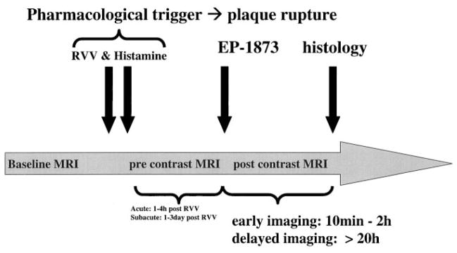
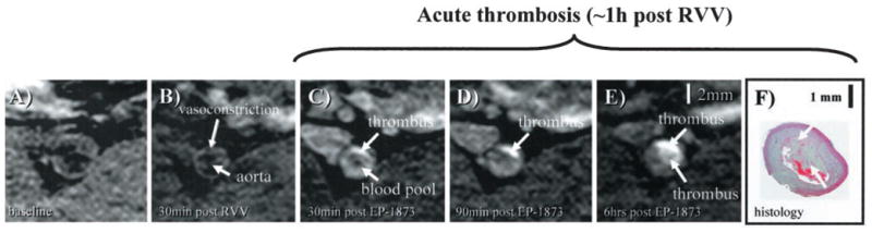
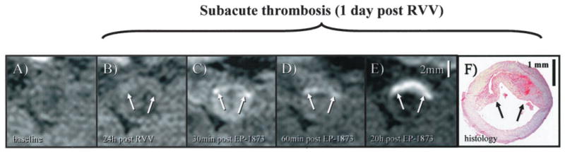
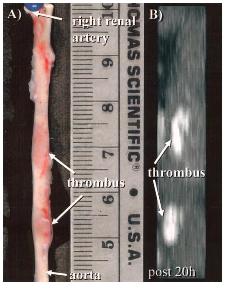
Methods and results: Fifteen male New Zealand White rabbits (weight, approximately 3.5 kg) were made atherosclerotic by feeding a high-cholesterol diet after endothelial aortic injury. Plaque rupture was then induced with the use of Russell's viper venom (RVV) and histamine. Subsequently, MRI of the subrenal aorta was performed before RVV, after RVV, and after EP-1873. Histology was performed on regions suggested by MRI to contain thrombus. Nine rabbits (60%) developed plaque rupture and thrombus, including 25 thrombi visually apparent on MRI as "hot spots" after injection of EP-1873. Histological correlation confirmed all 25 thrombi (100%), with no thrombi seen in the other regions of the aorta. In the remaining 6 rabbits (control) without plaque rupture, no thrombus was observed on the MR images or on histology.

Conclusions: We demonstrate the feasibility of in vivo "molecular" MRI for the detection of acute and subacute thrombosis using a novel fibrin-binding MRI contrast agent in an animal model of atherosclerosis and acute/subacute thrombosis. Potential clinical applications include thrombus detection in acute coronary syndromes and stroke.


Figures

Figure 1
Figure 1
Time course of in vivo MRI experiment of acute and subacute thrombosis with the use of an atherosclerotic New Zealand White rabbit model. After an 8-week high-cholesterol diet, baseline MRI of the subrenal aorta was performed. Subsequently, plaque rupture was induced with the use of RVV and histamine. The fibrin-binding peptide derivative EP-1873 was then intravenously administered ≈1 hour (imaging of acute thrombosis) or ≈20 hours (imaging of subacute thrombosis) after RVV and histamine. After intravenous injection of EP-1873, MRI was performed at 30 minutes and continued up to ≈20 hours. Subsequently, the animals were killed, and histological analyses were performed.
Figure 2
Figure 2
In vivo visualization of acute thrombosis after plaque rupture induced by RVV and histamine. A, No apparent thrombus is visible on baseline image. B, Thirty minutes after pharmacological triggering with RVV and histamine, vasoconstriction of the subrenal aorta can be observed without obvious signs of thrombosis. C, Thirty minutes after administration of EP-1873, a well-defined bright mass (arrow) can be observed, consistent with a mural thrombus. D, E, Thrombus increased in size and signal intensity over the subsequent 6 hours, consistent with ongoing thrombus formation. F, Histology demonstrates the corresponding thrombus (arrow) overlying a thickened atherosclerotic vessel wall.
Figure 3
Figure 3
In vivo visualization of subacute thrombosis after plaque rupture induced by RVV and histamine. A, On the pretrigger baseline image, no apparent thrombosis is visible. B, At 24 hours after trigger, a gray mass (arrow) overlying the vessel wall can be observed, suggestive of a mural thrombus. C, Thirty minutes after EP-1873 injection, a bright rim (arrow) becomes visible along the lumen-facing surface of this mass, which subsequently increased in size and signal intensity, as shown at 60 minutes (D). E, Twenty hours after EP-1873 injection, entire mass (arrow) appeared bright and well defined, suggesting that the fibrin-binding agent had completely penetrated the thrombus. Good agreement was found with thrombus (arrows) on histopathology (F).
Figure 4
Figure 4
A, Photograph demonstrates 2 well-delineated focal thrombi (arrows) in a harvested subrenal aorta. B, On corresponding targeted MIP, 2 bright thrombi (arrows) can be identified that correspond in both size and location with gross histological image in A.
Figure 5
Figure 5
A, Reformatted view of a coronal 3D data set shows subrenal aorta ≈20 hours after EP-1873 administration. Three well-delineated mural thrombi (arrows) can be observed, with good contrast between thrombus (numbered), arterial blood (dotted arrow), and vessel wall (dashed arrow). The in-plane view of the aorta allows simultaneous display of all thrombi, showing head, tail, length, and relative location. B to D, Corresponding cross-sectional views show good agreement with histopathology (E to G).
Figure 6
Figure 6
EP-1873 binding to rabbit fibrin in Tris-buffered saline with data fit best to Kd=3.5±0.15 μmol/L and 2.4±0.1 binding (bnd) sites.
Figure 7
Figure 7
Displacement of fibrin-bound paramagnetic EP-1873 in a rabbit carotid thrombus model after injection of nonparamagnetic Y-1873. A, Baseline image of carotid artery. B, Carotid thrombus after injection (1.45 μmol/kg) of fibrin-binding EP-1873. C to D, Displacement of paramagnetic EP-1873 by nonparamagnetic Y-1873 (14.5 μmol/kg), resulting in visually apparent decrease of thrombus signal.

References

    1. Fuster V, Badimon L, Badimon JJ, et al. The pathogenesis of coronary artery disease and the acute coronary syndromes. N Engl J Med. 1992;326:242–250. - PubMed
    1. Davies MJ, Thomas AC. Plaque fissuring: the cause of acute myocardial infarction, sudden ischaemic death, and crescendo angina. Br Heart J. 1985;53:363–373. - PMC - PubMed
    1. Grotta JC. Adding to the effectiveness of intravenous tissue plasminogen activator for treating acute stroke. Circulation. 2003;107:2769–2770. - PubMed
    1. de Korte CL, Sierevogel MJ, Mastik F, et al. Identification of atherosclerotic plaque components with intravascular ultrasound elastography in vivo: a Yucatan pig study. Circulation. 2002;105:1627–1630. - PubMed
    1. Abela GS, Eisenberg JD, Mittleman MA, et al. Detecting and differentiating white from red coronary thrombus by angiography in angina pectoris and in acute myocardial infarction. Am J Cardiol. 1999;83:94–97. A8. - PubMed

Publication types