. 2004 Mar;113(6):876-84.
doi: 10.1172/JCI19480.

Endothelin-1 regulates cardiac sympathetic innervation in the rodent heart by controlling nerve growth factor expression

Affiliations

Endothelin-1 regulates cardiac sympathetic innervation in the rodent heart by controlling nerve growth factor expression

Masaki Ieda et al. J Clin Invest. 2004 Mar.

Abstract

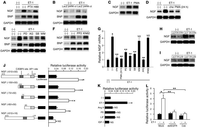
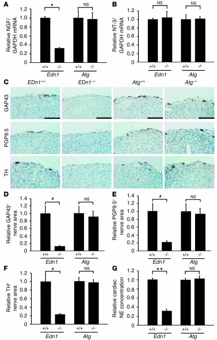
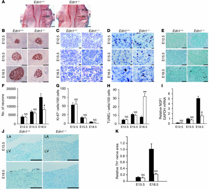
The cardiac sympathetic nerve plays an important role in regulating cardiac function, and nerve growth factor (NGF) contributes to its development and maintenance. However, little is known about the molecular mechanisms that regulate NGF expression and sympathetic innervation of the heart. In an effort to identify regulators of NGF in cardiomyocytes, we found that endothelin-1 specifically upregulated NGF expression in primary cultured cardiomyocytes. Endothelin-1-induced NGF augmentation was mediated by the endothelin-A receptor, Gibetagamma, PKC, the Src family, EGFR, extracellular signal-regulated kinase, p38MAPK, activator protein-1, and the CCAAT/enhancer-binding protein delta element. Either conditioned medium or coculture with endothelin-1-stimulated cardiomyocytes caused NGF-mediated PC12 cell differentiation. NGF expression, cardiac sympathetic innervation, and norepinephrine concentration were specifically reduced in endothelin-1-deficient mouse hearts, but not in angiotensinogen-deficient mice. In endothelin-1-deficient mice the sympathetic stellate ganglia exhibited excess apoptosis and displayed loss of neurons at the late embryonic stage. Furthermore, cardiac-specific overexpression of NGF in endothelin-1-deficient mice overcame the reduced sympathetic innervation and loss of stellate ganglia neurons. These findings indicate that endothelin-1 regulates NGF expression in cardiomyocytes and plays a critical role in sympathetic innervation of the heart.


Figures

Figure 1
Figure 1
Specific augmentation of NGF expression by ET-1 in cardiomyocytes. (A) Gene expression of four NGF alternatively spliced transcripts (a–d) in murine heart (H), brain (BR), and submaxillary gland (S) was determined by RT-PCR. The number of PCR cycles is 35. m, marker. (B) Cardiomyocytes were stimulated with ET-1, angiotensin II (Ang II), phenylephrine (PE), LIF, or IGF-1 for 2 hours. Northern blot analysis for NGF was performed. (C) Stimulation of cardiomyocytes with ET-1 for specified time intervals. (D) Stimulation of cardiomyocytes with various concentrations of ET-1. (E) Cardiomyocytes were pretreated with BQ123 (BQ) or TAK044 (TAK) for 30 minutes; then RNA was isolated 2 hours after ET-1 stimulation. (F) Induction of NGF expression by ET-1 in cardiomyocytes (CM), but not in cardiac fibroblasts (CF). Results similar to those shown in A–F were obtained in four separate experiments. (G) NGF protein levels in conditioned medium. Augmentation of NGF protein was inhibited by pretreatment with BQ123. (n = 4.) (H) Secretion of NGF by cardiomyocytes was not induced by angiotensin II, phenylephrine, LIF, or IGF-1 (n = 4). *P < 0.001. NS, not significant vs. control.
Figure 2
Figure 2
The ET-1/ETA receptor augments NGF expression via Giβγ, PKC, EGFR, ERK, p38MAPK, and AP-1 and C/EBPδ elements. (A) Preincubation of cardiomyocytes with either PTX or H89. NGF mRNA expression was determined 2 hours after ET-1 stimulation. (B) Cardiomyocytes were pretreated with LacZ or βARK-ct to inhibit the function of Giβγ, and stimulated with ET-1. βARK-ct attenuated ET-1–induced NGF expression, but not BNP. (C and D) Stimulation with PMA (a PKC activator) for 2 hours augmented NGF expression. In contrast, pretreatment with chelerythrine (che; a PKC inhibitor) for 30 minutes or PMA for 24 hours inhibited ET-1–induced NGF expression. (E and F) Pretreatment with PD98059 (PD; an MAPK inhibitor), AG1478 (AG; an EGFR inhibitor), SB203580 (SB; a p38MAPK inhibitor), or PP2 (an Src family inhibitor), but not with wortmannin (WM; a PI3K inhibitor) or KN62 (a calmodulin kinase II/Iv inhibitor) attenuated ET-1–induced NGF mRNA expression. BNP was affected only with PD98059 pretreatment. (G) The results of the densitometry of four separate experiments are shown. *P < 0.001 vs. control; **P < 0.01 vs. ET-1 alone. NS, not significant vs. ET-1 alone. (H and I) Cardiomyocytes were pretreated with DN-ERK or DN-p38MAPK. (J and K) Identification of ET-1–responsive elements in the NGF promoter using luciferase assay. Black bars, control; white bars, ET-1 stimulation (n = 4). (L) Specific negative regulatory plasmid of the EGFR (533delEGFR) or the Src family (Csk) inhibited NGF transcription (n = 4). *P < 0.001, **P < 0.01, #P < 0.05 vs. relative control. NS, not significant.
Figure 3
Figure 3
ET-1 causes NGF-mediated differentiation of PC12 cells. (A) PC12 cell morphology was observed after incubation for 3 days in mock medium or medium conditioned with unstimulated cardiomyocytes (U-CM) or ET-1–stimulated cardiomyocytes (ET-CM). Medium conditioned with ET-1–stimulated cardiomyocytes strongly induced neurite extension in PC12 cells compared with medium conditioned with unstimulated cardiomyocytes. (B) Percentage of differentiated cells in A (n = 4). (C) PC12 cells were pretreated with anti-NGF blocking antibody for 30 minutes, then incubated with the conditioned medium (n = 4). (D) PC12 cells transfected with LacZ were cocultured with cardiomyocytes, then stimulated with ET-1 or ET-1 plus anti-NGF blocking antibody for 3 days. PC12 cells were identified using X-gal staining. (E) Percentage of differentiated cells in D (n = 4). (F) NGF protein levels in medium conditioned with Edn1+/+ or Edn1–/– cardiomyocytes were measured by ELISA (n = 4). *P < 0.0001; **P < 0.001; #P < 0.01. Scale bar: 100 μm.
Figure 4
Figure 4
Disruption of ET-1, but not of angiotensinogen, reduces NGF expression, sympathetic nerve density, and norepinephrine concentration in murine hearts. (A) NGF expression in Edn1+/+, Edn1–/–, Atg+/+, and Atg–/– hearts at E18.5 was determined by quantitative RT-PCR (n = 10). (B) Neurotrophin-3 (NT-3) expression in the heart was measured by quantitative RT-PCR. The same reverse transcription products used in A were analyzed. (n = 10.) (C) Immunostaining for GAP43, PGP9.5, and TH in the heart. Nerves were restricted to the epicardium in both genotypes, and levels of GAP43, PGP9.5, and TH were lower in Edn1–/– mice, but not in Atg–/– mice, compared with WT littermates. (D–F) The immunopositive nerve areas for GAP43, PGP9.5, and TH were determined using NIH Image (n = 8). (G) Cardiac norepinephrine (NE) concentrations were measured by HPLC (n = 10). *P < 0.0001; **P < 0.005; #P < 0.01. NS, not significant. Scale bar: 100 μm.
Figure 5
Figure 5
Edn1–/– embryos display a loss of SG neurons due to excess apoptosis. (A) Edn1+/+ and Edn1–/– whole-mount embryos at E12.5 were immunostained with anti-TH antibody. SCG, superior cervical ganglion; fl, forelimb. Similar results were obtained from four separate experiments. (B–E) TH immunostaining, cresyl violet staining (CV), Ki-67 immunostaining, and TUNEL staining of Edn1+/+ and Edn1–/– SG at E12.5, E15.5, and E18.5 at the same level of section. Note that at E18.5, Edn1–/– SG were considerably smaller than Edn1+/+ SG and increased apoptosis was detected. (F–H) Time course of the number of neurons, Ki-67+ cells per 100 neurons, and TUNEL+ cells per 1,000 neurons in SG was shown (n = 5). (I) Time course of NGF expression in Edn1+/+ and Edn1–/– hearts was determined by quantitative RT-PCR (n = 3). (J and K) Immunostaining for TH in the heart of Edn1+/+ and Edn1–/– embryos at E15.5 and E18.5. TH-immunopositive nerve fibers were slightly detected from E15.5. LA, left atrium; LV, left ventricle. The immunopositive nerve areas for TH were determined using NIH Image. (n = 4.) *P < 0.001; **P < 0.01; #P < 0.05. NS, not significant vs. relative control. Scale bar: 500 μm (A), 100 μm (B and J), 10 μm (C and D), 50 μm (E). Black bars, Edn1+/+; white bars, Edn1–/–.
Figure 6
Figure 6
Cardiac-specific overexpression of NGF overcomes the defects of the cardiac sympathetic nervous system in Edn1–/– mice. (A) NGF expression in Edn1+/+, Edn1–/–, and Edn1–/–/MHC-NGF hearts is shown (n = 6). The reduced NGF expression in the Edn1–/– heart was completely overcome by cardiac-specific overexpression of NGF. (B) Immunostaining for TH in the hearts of Edn1+/+, Edn1–/–, and Edn1–/–/MHC-NGF mice. Scale bar: 100 μm. (C) The immunopositive nerve areas for TH were quantitated (n = 6). (D) The cardiac norepinephrine (NE) concentration was increased in Edn1–/–/MHC-NGF mice compared with Edn1–/– mice (n = 6). (E) TH immunostaining, cresyl violet staining (CV), Ki-67 immunostaining, and TUNEL staining of Edn1+/+, Edn1–/–, and Edn1–/–/MHC-NGF SG at E18.5 at the same level of section. Note that the reduction of the size of SG and the increase in TUNEL+ cells in Edn1–/– mice were completely reversed in Edn1–/–/MHC-NGF mice. (F and G) The number of neurons and the number of TUNEL+ cells per 1,000 neurons in each SG are shown (n = 3–6). *P < 0.0001; **P < 0.01; #P < 0.05. TG, transgenic. Scale bar: 100 μm (TH), 50 μm (TUNEL), 10 μm (CV and Ki-67).

Comment in

  • Sculpting organ innervation.
    Hempstead BL. Hempstead BL. J Clin Invest. 2004 Mar;113(6):811-3. doi: 10.1172/JCI21309. J Clin Invest. 2004. PMID: 15067313 Free PMC article.

Similar articles

Cited by

References

    1. Loring JF, Erickson CA. Neural crest cell migratory pathways in the trunk of the chick embryo. Dev. Biol. 1987;121:220–236. - PubMed
    1. Snider WD. Functions of the neurotrophins during nervous system development: what the knockouts are teaching us. Cell. 1994;77:627–638. - PubMed
    1. Lockhart ST, Turrigiano GG, Birren SJ. Nerve growth factor modulates synaptic transmission between sympathetic neurons and cardiac myocytes. J. Neurosci. 1997;17:9573–9582. - PMC - PubMed
    1. Heumann R, Korsching S, Scott J, Thoenen H. Relationship between levels of nerve growth factor (NGF) and its messenger RNA in sympathetic ganglia and peripheral target tissues. EMBO J. 1984;3:3183–3189. - PMC - PubMed
    1. Brennan C, Rivas-Plata K, Landis SC. The p75 neurotrophin receptor influences NT-3 responsiveness of sympathetic neurons in vivo. Nat. Neurosci. 1999;2:699–705. - PubMed

Publication types