. 2004 Jun 4;279(23):24197-202.
doi: 10.1074/jbc.M402452200. Epub 2004 Apr 6.

Structural and ligand recognition characteristics of an acetylcholine-binding protein from Aplysia californica

Affiliations

Structural and ligand recognition characteristics of an acetylcholine-binding protein from Aplysia californica

Scott B Hansen et al. J Biol Chem. .

Abstract

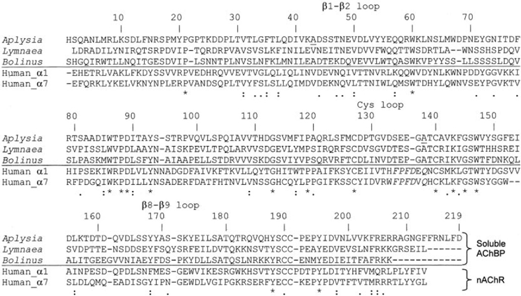
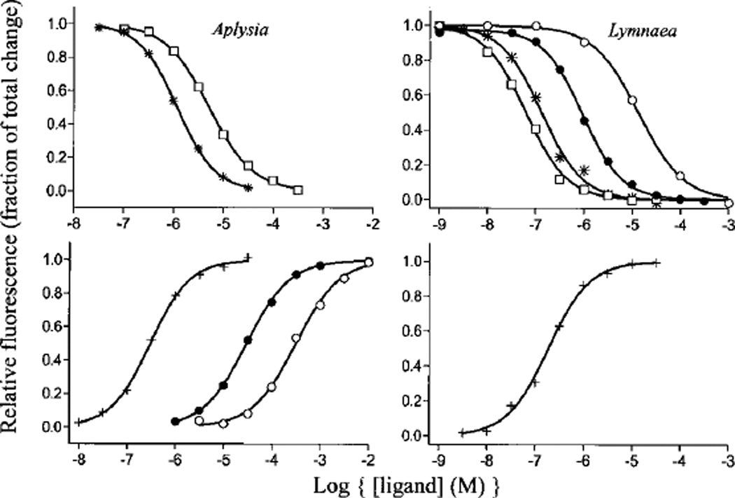
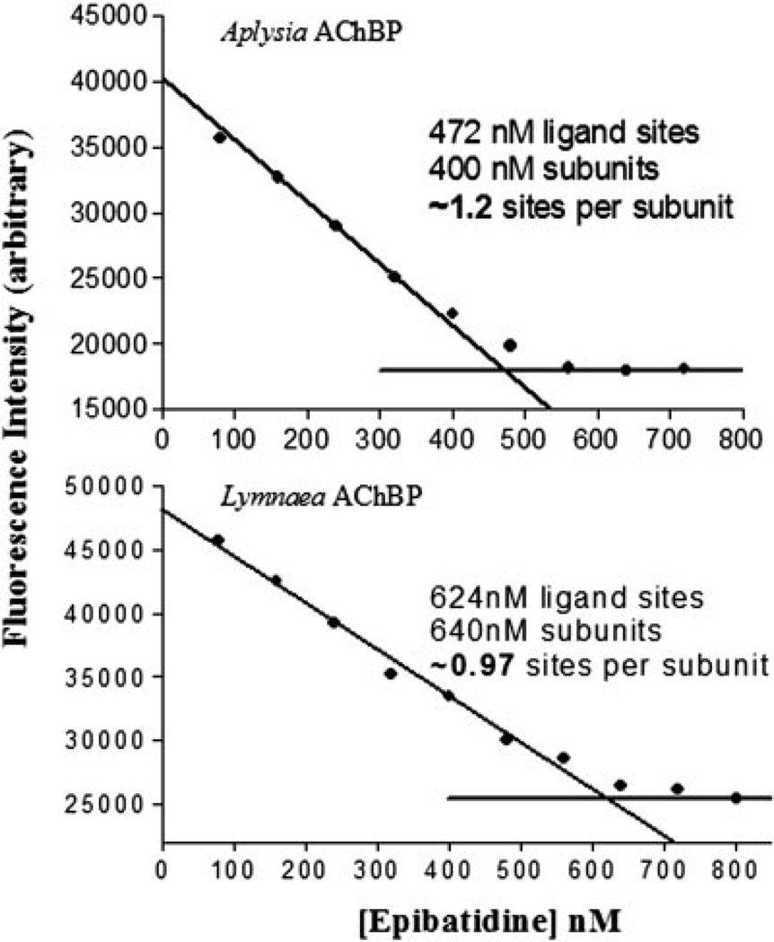
We generated an acetylcholine-binding protein from Aplysia californica by synthesis of a cDNA found in existing data bases and its expression in mammalian cell culture. Its subunit assembly and ligand recognition behavior were compared with the binding protein previously derived from Lymnaea stagnalis. The secreted proteins were purified by elution from columns of attached antibodies directed to the FLAG epitope encoded in the expression construct. Although the sequences of the two proteins from marine and fresh water mollusks exhibit the characteristic features of the extracellular domain of the nicotinic receptor, they only possess 33% amino acid identity. Both assemble as stable pentamers with five binding sites per pentamer, yet they show distinguishing features of stability and sensitivity to epitope tag placement. Both proteins exhibit changes in tryptophan fluorescence upon ligand binding; however, the magnitude of the changes differs greatly. Moreover, certain ligands show marked differences in dissociation constants for the two proteins and can be regarded as distinguishing or signature ligands. Hence, the two soluble proteins from mollusks, which can be studied by a variety of physical methods, become discrete surrogate proteins for the extracellular domains of distinct subtypes of nicotinic acetylcholine receptors.


Figures

Fig. 1
Fig. 1. Protein sequence alignment
Three soluble binding proteins from L. stagnalis, A. californica, and Bolinus truncatus (22) are aligned with the first 210 amino acid residues of human nicotinic acetylcholine receptor α1 and α7 subunits. The numbering corresponds to Aplysia, beginning with the first synthesized residue in the cDNA sequence and a probable start site based on consensus sequences. The asterisks indicate identity among the receptor family, whereas colons and periods indicate limited conservation in the series. Underscored alanines 43 and 138 were found to be valines in a sensory cell Aplysia cDNA library.
Fig. 2
Fig. 2. AChBP cDNA constructs used to characterize the acetylcholine binding protein
PPT, a preprotrypsin leader peptide. Dark shading indicates a purification tag (3× or 1×, FLAG epitope; H, 6× histidine). Equivalent constructs were made with both Aplysia and Lymnaea. The numbers indicate the respective amino acid residues in the assembled gene products coming from the sequences in Fig. 1.
Fig. 3
Fig. 3. Fast protein liquid chromatography of AChBPs
A, non-His-tagged A-AChBP (II); B, non-His-tagged L-AChBP (II); C and D, His-tagged A-AChBP (I). The Roman numerals designate the constructs in Fig. 2. C, ~50% aggregated protein; D, mostly properly assembled pentamer with a small shoulder of aggregate. The arrows indicate an apparentMr 190,000 pentamer peak, and vertical lines indicate the void volume. Purified AChBP (0.1 mg/ml) in 20 mm Tris-HCl, 150 mm NaCl, pH 7.4, was applied to a Superdex 200 size exclusion column.
Fig. 4
Fig. 4. SDS-PAGE electrophoresis of the purified acetylcholine-binding proteins
Lanes 1–4, Lymnaea proteins; lanes 5–8, Aplysia proteins. Size and glycosylation are distinct among tags and seem roughly similar between species. Purified protein (1.5 µg) was spotted on each lane and run on 16% acrylamide gels. The Roman numerals denote the constructs described in the legend to Fig. 2.
Fig. 5
Fig. 5. Analytical ultracentrifugation of Aplysia AChBP
Samples of 100 µg/ml A-AChBP in (20 mm Tris-HCl, 150 mm NaCl, pH 7.4) was sedimented at 12,000 rpm until a constant profile was established. The curve is fit to a molecular weight of 153,000 with the other variables fixed as described under “Materials and Methods.”
Fig. 6
Fig. 6. pH Stability of the AChBPs
Samples of 0.5 nm AChBP binding sites were incubated with 20 nm [3H]epibatidine for 1 h and monitored using a scintillation proximity assay. The pH was varied between 5.0 and 11 using a 100 mm phosphate/pyrophosphate buffer. ○, L-AChBP; ●, A-AChBP.
Fig. 7
Fig. 7. Equilibrium ligand binding to AChBPs
Ligand binding was monitored in a 96-well fluorescent plate reader. Samples were excited at 280 nm, and intrinsic tryptophan fluorescence was emission-monitored at 340 nm. ○, carbachol; ●, acetylcholine; +, gallamine; *, dansylcholine C6; □, nicotine). Dissociation constants from experiments such as this are shown in Table II. Left panels, Aplysia AChBP; right panels, Lymnaea AChBP.
Fig. 8
Fig. 8. Titration of ligand stoichiometry
Using excess ligand binding sites over Kd and monitoring intrinsic tryptophan fluorescence quenching of AChBP at 340 nm, binding site titration with [3H]epibatidine was used to estimate the total number of binding sites. Saturation occurs at ~5 sites/pentamer. Top, Aplysia AChBP; bottom, Lymnaea AChBP.

References

    1. Corringer PJ, Le Novere N, Changeux JP. Annu. Rev. Pharmacol. Toxicol. 2000;40:431–458. - PubMed
    1. Karlin A. Nat. Rev. Neurosci. 2002;3:102–114. - PubMed
    1. Sixma TK, Smit AB. Annu. Rev. Biophys. Biomol. Struct. 2003;32:311–334. - PubMed
    1. Chang CC, Lee CY. Arch. Int. Pharmacodyn. Ther. 1963;144:241–257. - PubMed
    1. Lee CY. Showa Igakkai Zasshi. 1963;23:221–229. - PubMed

Publication types

MeSH terms

LinkOut - more resources