

Hepatic progenitor cells in human liver cirrhosis: immunohistochemical, electron microscopic and immunofluorencence confocal microscopic findings
- PMID: 15069727
- PMCID: PMC4656362
- DOI: 10.3748/wjg.v10.i8.1208
Hepatic progenitor cells in human liver cirrhosis: immunohistochemical, electron microscopic and immunofluorencence confocal microscopic findings
Abstract
Aim: To investigate whether hepatic progenitor cells (HPC), that reveal the features of oval cells in rodents and small epithelial cells (SEC) in certain human liver disease, were also found in human liver cirrhosis (HLC).
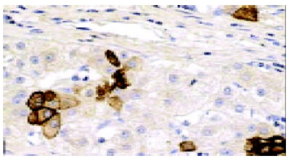
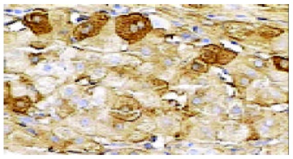
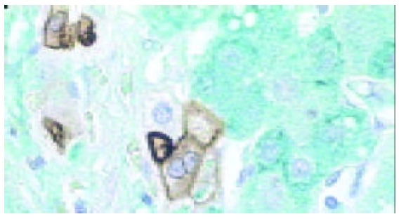
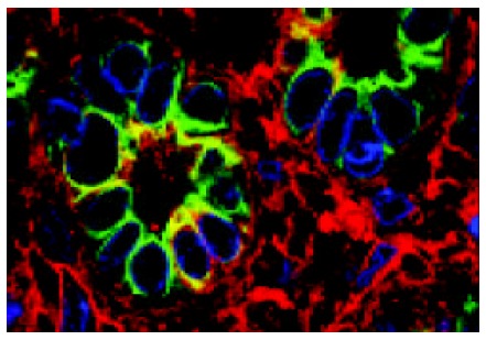
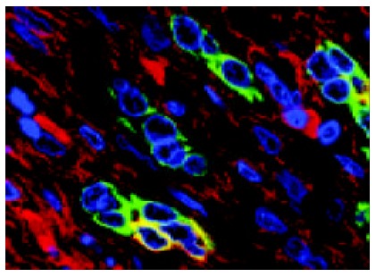
Methods: Surgical liver specimens from 20 cases of hepatitis B virus-positive HLC (15 cases containing hepatocellular carcinoma) were investigated by light microscopic immunohistochemistry (LM-IHC). Among them specimens from 15 cases were investigated by electron microscopy (EM) and those from 5 cases by immunofluorencence confocal laser scanning microscopy (ICLSM). Antibodies against cytokeratin 7 and albumin were used and single and/or double labelling were performed respectively.
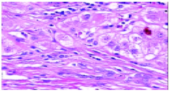
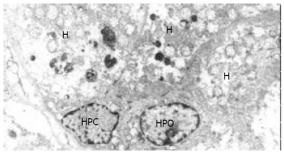
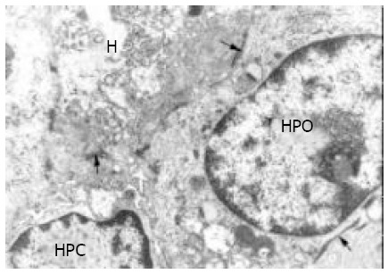
Results: LM-IHC showed that at the margins of regenerating nodules and in the fibrous septae, a small number of cells in the proliferating bile ductules were positive for CK7 and albumin. At the EM level these HPC were morphologically similar to the SEC described previously, and also similar to the oval cells seen in experimental hepatocarcinogenesis. They were characterized by their small size, oval shape, a high nucleus/cytoplasm ratio, a low organelle content in cytoplasm, and existence of tonofilaments and intercellular junctions. ICLSM revealed that HPC expressed both cytokeratin 7 and albumin.
Conclusion: HPC with ultrastructural and immunophenotypical features of oval cells, i.e., hepatic stem cell-like cells as noted in other liver diseases, were found in HLC. These findings further support the hypothesis that bipotent hepatic stem cells, that may give rise to biliary epithelial cells and hepatocytes, exist in human livers.
Figures
References
-
- Forbes S, Vig P, Poulsom R, Thomas H, Alison M. Hepatic stem cells. J Pathol. 2002;197:510–518. - PubMed
-
- Yin L, Lynch D, Ilic Z, Sell S. Proliferation and differentiation of ductular progenitor cells and littoral cells during the regeneration of the rat liver to CCl4/2-AAF injury. Histol Histopathol. 2002;17:65–81. - PubMed
Publication types
MeSH terms
LinkOut - more resources
Full Text Sources
Medical
Research Materials
