

Spatiotemporal dynamics of word processing in the human cortex
- PMID: 15070488
- PMCID: PMC3746799
- DOI: 10.1177/1073858403261018
Spatiotemporal dynamics of word processing in the human cortex
Abstract
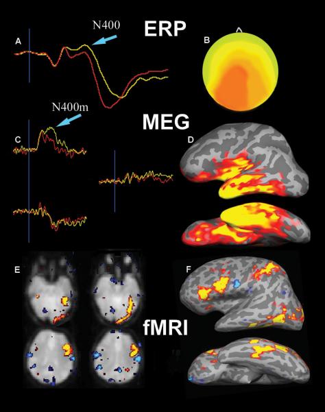
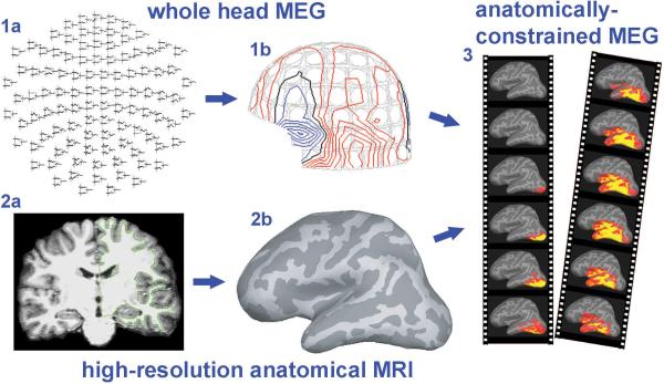
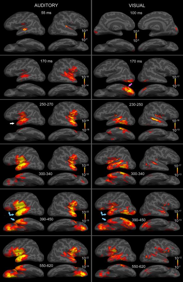
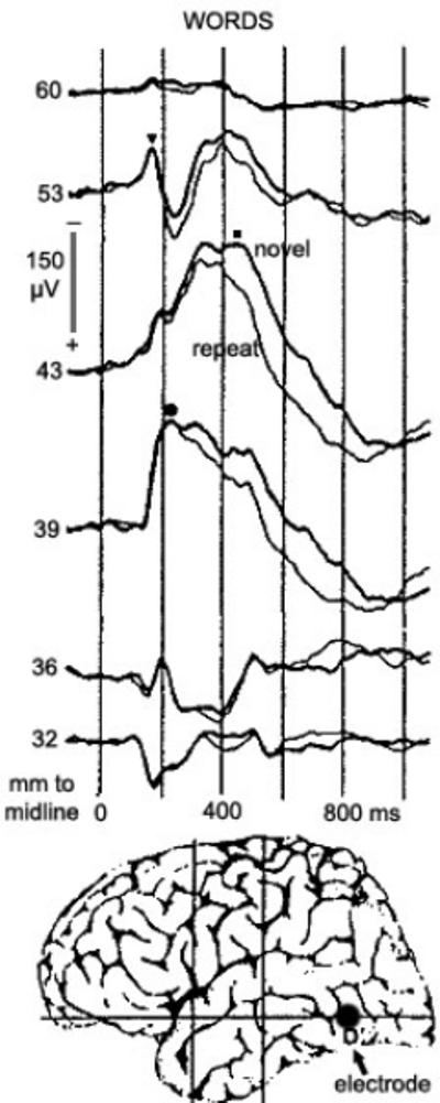
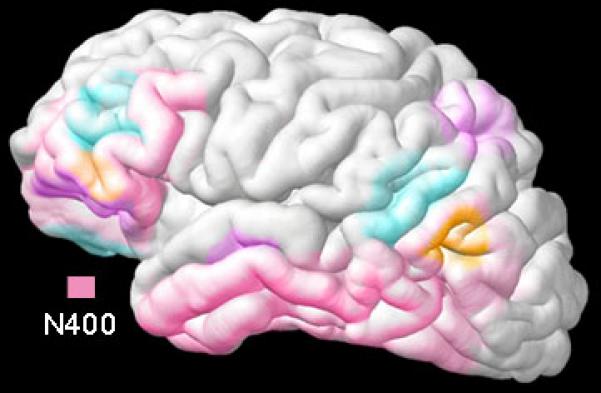
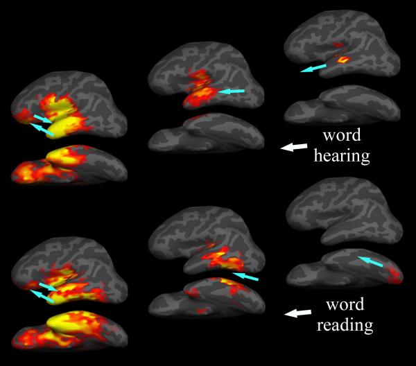
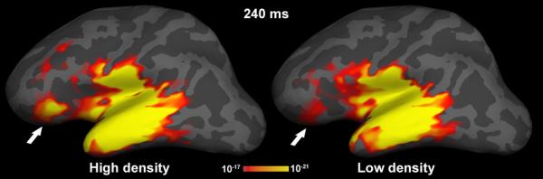
Understanding language relies on concurrent activation of multiple areas within a distributed neural network. Hemodynamic measures (fMRI and PET) indicate their location, and electromagnetic measures (magnetoencephalography and electroencephalography) reveal the timing of brain activity during language processing. Their combination can show the spatiotemporal characteristics (where and when) of the underlying neural network. Activity to written and spoken words starts in sensory-specific areas and progresses anteriorly via respective ventral ("what") processing streams toward the simultaneously active supramodal regions. The process of understanding a word in its current context peaks about 400 ms after word onset. It is carried out mainly through interactions of the temporal and inferior prefrontal areas on the left during word reading and bilateral temporo-prefrontal areas during speech processing. Neurophysiological evidence suggests that lexical access, semantic associations, and contextual integration may be simultaneous as the brain uses available information in a concurrent manner, with the final goal of rapidly comprehending verbal input. Because the same areas may participate in multiple stages of semantic or syntactic processing, it is crucial to consider both spatial and temporal aspects of their interactions to appreciate how the brain understands words.
Figures
Similar articles
-
Spatiotemporal dynamics of modality-specific and supramodal word processing.Neuron. 2003 May 8;38(3):487-97. doi: 10.1016/s0896-6273(03)00197-1. Neuron. 2003. PMID: 12741994 Free PMC article.
-
Spatiotemporal dynamics of neural language processing: an MEG study using minimum-norm current estimates.Neuroimage. 2003 Oct;20(2):1020-5. doi: 10.1016/S1053-8119(03)00356-2. Neuroimage. 2003. PMID: 14568471 Clinical Trial.
-
Balancing Prediction and Sensory Input in Speech Comprehension: The Spatiotemporal Dynamics of Word Recognition in Context.J Neurosci. 2019 Jan 16;39(3):519-527. doi: 10.1523/JNEUROSCI.3573-17.2018. Epub 2018 Nov 20. J Neurosci. 2019. PMID: 30459221 Free PMC article.
-
Words in the brain's language.Behav Brain Sci. 1999 Apr;22(2):253-79; discussion 280-336. Behav Brain Sci. 1999. PMID: 11301524 Review.
-
[Anatomic and topographic models of the cerebral areas that activates during the linguistic functions].Rev Neurol. 2008 Dec 16-31;47(12):653-8. Rev Neurol. 2008. PMID: 19085883 Review. Spanish.
Cited by
-
Head position in the MEG helmet affects the sensitivity to anterior sources.Neurol Clin Neurophysiol. 2004 Nov 30;2004:30. Neurol Clin Neurophysiol. 2004. PMID: 16012659 Free PMC article.
-
Spatiotemporal dynamics of bilingual word processing.Neuroimage. 2010 Feb 15;49(4):3286-94. doi: 10.1016/j.neuroimage.2009.12.009. Epub 2009 Dec 11. Neuroimage. 2010. PMID: 20004256 Free PMC article.
-
Topographic mapping of a hierarchy of temporal receptive windows using a narrated story.J Neurosci. 2011 Feb 23;31(8):2906-15. doi: 10.1523/JNEUROSCI.3684-10.2011. J Neurosci. 2011. PMID: 21414912 Free PMC article.
-
Reading front to back: MEG evidence for early feedback effects during word recognition.Cereb Cortex. 2014 Mar;24(3):817-25. doi: 10.1093/cercor/bhs365. Epub 2012 Nov 21. Cereb Cortex. 2014. PMID: 23172772 Free PMC article.
-
MEG Theta during Lexico-Semantic and Executive Processing Is Altered in High-Functioning Adolescents with Autism.Cereb Cortex. 2021 Jan 5;31(2):1116-1130. doi: 10.1093/cercor/bhaa279. Cereb Cortex. 2021. PMID: 33073290 Free PMC article.
References
-
- Bar M. A cortical mechanism for triggering top-down facilitation in visual object recognition. J Cogn Neurosci. 2003;15(4):600–9. - PubMed
-
- Besson M, Schon D. Comparison between language and music. Ann N Y Acad Sci. 2001;930:232–58. - PubMed
-
- Brownell HH, Simpson TL, Bihrle AM, Potter HH, Gardner H. Appreciation of metaphoric alternative word meanings by left and right brain-damaged patients. Neuropsychologia. 1990;28(4):375–83. - PubMed
-
- Buckner RL, Koutstaal W, Schacter DL, Rosen BR. Functional MRI evidence for a role of frontal and inferior temporal cortex in amodal components of priming. Brain. 2000;123(Pt 3):620–40. - PubMed
Publication types
MeSH terms
Grants and funding
LinkOut - more resources
Full Text Sources
