. 2004 Apr 15;103(8):3029-37.
doi: 10.1182/blood-2003-10-3417. Epub 2004 Jan 8.

Tissue factor-factor VIIa-specific up-regulation of IL-8 expression in MDA-MB-231 cells is mediated by PAR-2 and results in increased cell migration

Affiliations

Tissue factor-factor VIIa-specific up-regulation of IL-8 expression in MDA-MB-231 cells is mediated by PAR-2 and results in increased cell migration

Gertrud M Hjortoe et al. Blood. .

Abstract

Tissue factor (TF), the cellular receptor for factor VIIa (FVIIa), besides initiating blood coagulation, is believed to play an important role in tissue repair, inflammation, angiogenesis, and tumor metastasis. Like TF, the chemokine interleukin-8 (IL-8) is shown to play a critical role in these processes. To elucidate the potential mechanisms by which TF contributes to tumor invasion and metastasis, we investigated the effect of FVIIa on IL-8 expression and cell migration in a breast carcinoma cell line, MDA-MB-231, a cell line that constitutively expresses abundant TF. Expression of IL-8 mRNA in MDA-MB-231 cells was markedly up-regulated by plasma concentrations of FVII or an equivalent concentration of FVIIa (10 nM). Neither thrombin nor other proteases involved in hemostasis were effective in stimulating IL-8 in these cells. Increased transcriptional activation of the IL-8 gene is responsible for increased expression of IL-8 in FVIIa-treated cells. PAR-2-specific antibodies fully attenuated TF-FVIIa-induced IL-8 expression. Additional in vitro experiments showed that TF-FVIIa promoted tumor cell migration and invasion, active site-inactivated FVIIa, and specific antibodies against TF, PAR-2, and IL-8 inhibited TF-FVIIa-induced cell migration. In summary, the studies described herein provide insight into how TF may contribute to tumor invasion.


Figures

Figure 1
Figure 1. FVIIa-induced IL-8 expression in breast carcinoma cells
Quiescent monolayers of MDA-MB-231 cells were treated with varying concentrations of FVIIa (A-C), a plasma concentration (10 nM) of zymogen FVII or FVIIa (D), or varying concentrations of active site-inactivated FVIIa (ASIS/FFR-FVIIa), followed by 10 nM FVIIa (E). (F) Cells were pretreated with anti-TF IgG or control IgG (100 μg/mL) for 45 minutes before FVIIa (10 nM) was added to the cells. Unless otherwise specified, cells were treated with FVIIa for 75 minutes at 37°C, and total RNA was isolated and subjected to Northern blot analysis. (A, D-F) Representative Northern blot analysis of IL-8 mRNA; (B) quantitative data from such experiments. (C) IL-8 antigen levels in overlying conditioned media of MDA-MB-231 cells treated with varying concentrations of FVIIa. Error bars indicate SEM from 2 to 3 experiments.
Figure 2
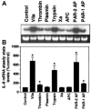
Figure 2. Effect of FVIIa and other agonists on the induction of IL-8 mRNA
Quiescent monolayers of MDA-MB-231 cells were treated with FVIIa (50 nM), thrombin (10 nM), plasmin (50 nM), trypsin (50 nM), FXa (50 nM), APC (50 nM), PAR-2–specific peptide agonist SLIGKV (25 μM) and PAR-1–specific peptide agonist TFLLRN (25 μM) for 75 minutes. Total RNA was analyzed for IL-8 expression by Northern blot analysis. (A) Representative autoradiograph. (B) Quantitative data (mean ± SEM, n = 4 to 7). *Value significantly higher (P < .05) than the control value.
Figure 3
Figure 3. Expression and functional activity of PAR-1 and PAR-2 in MDA-MB-231 cells
(A) MDA-MB-231 cells were probed with anti–PAR-1 (ATAP2) or anti–PAR-2 (SAM11) monoclonal antibodies, followed by FITC-labeled secondary antibody. FITC-labeled cells were analyzed by flow cytometry. Solid lines represent background fluorescence (control IgG), whereas dotted lines represent fluorescence shift attributable to PAR expression. (B) Intracellular calcium fluxes in response to PAR-1– and PAR-2–specific peptide agonists, or FVIIa. Fluo-4–loaded cells were exposed to a control medium, PAR-1–, or PAR-2–specific peptide agonists (50 μM) or FVIIa (100 nM). The resultant change in fluorescence at 520 nm after excitation at 485 nm is presented as relative fluorescence change compared with basal level fluorescence measured before the addition of compounds.
Figure 4
Figure 4. PAR-2 antibody inhibits FVIIa-induced IL-8 mRNA induction
MDA-MB-231 cells were pretreated with control rabbit IgG (500 μg/mL), rabbit anti PAR-2 IgG (500 μg/mL), or monoclonal antibodies against PAR-1 (10 μg/mL ATAP2 plus 25 μg/mL WEDE15) for 1 hour before they were stimulated with FVIIa (50 nM) for 75 minutes. Total RNA was analyzed by Northern blot analysis for IL-8 mRNA, and the hybridization signals were quantitated. Data shown in the figure represent mean ± SEM from 3 to 6 experiments.
Figure 5
Figure 5. Effect of FXa on FVIIa-induced IL-8 expression
MDA-MB-231 cells were stimulated for 75 minutes with varying concentrations of FVIIa in the presence and absence of FX (175 nM). As controls, cells were stimulated with FX (175 nM) or FXa (175 nM). Induction of IL-8 mRNA was analyzed by Northern blot analysis and quantitated using a PhosphorImager (mean ± SEM, n = 4). NS indicates not statistically significant (P = .3).
Figure 6
Figure 6. Effect of FVIIa on IL-8 mRNA stability and gene transcription
(A) MDA-MB-231 cells were first treated with a control vehicle or FVIIa (50 nM) for 75 minutes. Then 10 μg/mL actinomycin D was added to inhibit RNA synthesis. Total RNA was harvested at indicated times after the addition of actinomycin D and was subjected to Northern blot analysis using IL-8 probe. IL-8 mRNA levels measured 10 minutes after the addition of actinomycin D were taken as 100% (mean ± SEM, n = 3). (B) Nuclei were isolated from unstimulated MDA-MB-231 cells or cells stimulated with FVIIa (50 nM) for 1 hour. Two identical blots containing β-actin and IL-8 DNAs were hybridized with equal amounts of labeled transcripts of nuclear RNA. (C) Quantitative representation of the data shown in panel B (mean values of 2 experiments).
Figure 7
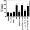
Figure 7. Effect of FVIIa and other agonists on cancer cell migration: involvement of PAR-2 and IL-8 in FVIIa-induced cell migration
MDA-MB-231 cells were placed in the upper well, and various concentrations of FVIIa (A) or other agonists (B) or FVIIa and antibodies against IL-8 or PAR-2 (C) were added to the lower well. In additional experiments, ASIS or anti-TF IgG was included with FVIIa in the lower well (A). The number of cells that migrated to the underside of the membrane in 20 hours at 37°C was determined as described in “Materials and methods” (mean ± SEM, n = 3 to 5). Concentrations of various reagents were: FVIIa, 50 nM (unless specified otherwise); ASIS, 500 nM; anti-TF IgG, 100 μg/mL; thrombin, 10 nM; trypsin, 10 nM; plasmin, 50 nM; PAR-1 AP, 50 μM; PAR-2 AP, 50 μM; rabbit antihuman IL-8 IgG, 50 μg/mL; rabbit anti–PAR-2 IgG, 500 μg/mL; control IgG, 50 μg/mL (1) or 500 μg/mL (2); and IL-8, 100 ng/mL. *Difference in value is statistically significant (P < .05) from the value obtained in control treatment (panel B) or corresponding control IgG (panel C).
Figure 8
Figure 8. Effect of FVIIa on tumor cell invasion
MDA-MB-231 cells were placed on top of a Matrigel barrier. FVIIa and other agonists were added to a lower well that contained NIH3T3 cell–conditioned media. At the end of a 48-hour incubation period at 37°C, the number of cells that migrated across the Matrigel barrier to the underside of the membrane was determined. Concentrations of various reagents used were: FVIIa, 50 nM; thrombin, 10 nM; PAR-2 AP, 50 μM; rabbit antihuman IL-8 IgG, 50 μg/mL; rabbit anti–PAR-2 IgG, 500 μg/mL; control IgG, 50 μg/mL (1) or 500 μg/Ml (2). Mean ± SEM, n = 3 to 5. *Inhibition was statistically significant.

References

    1. Trousseau A. Phlegmasia alba dolens: clinique medicale de l’Hotel-Dieu de Paris. The New Sydenham Society. 1865;3:654–712.
    1. Sack GH, Levin J, Bell WR. Trousseau’s syndrome and other manifestations of chronic disseminated coagulopathy in patients with neoplasms: clinical, pathophysiologic, and therapeutic features. Medicine. 1977;56:1–37. - PubMed
    1. Lieberman JS, Borrero J, Urdaneta E, Wright IS. Thrombophlebitis and cancer. JAMA. 1961;177:542–545. - PubMed
    1. Fisher MM, Hochberg LA, Wilensky ND. Recurrent thrombophlebitis on obscure malignant tumors of lung: report of four cases. JAMA. 1951;147:1213–1216. - PubMed
    1. Perlow S, Daniels JL. Venous thrombosis and obscure visceral carcinoma: report of ten cases. Arch Intern Med. 1956;97:184–188. - PubMed

Publication types

MeSH terms