. 2004 Mar 30;101(13):4361-6.
doi: 10.1073/pnas.0305705101. Epub 2004 Mar 2.

Mitochondrial DNA ligase in Crithidia fasciculata

Affiliations

Mitochondrial DNA ligase in Crithidia fasciculata

Krishna Murari Sinha et al. Proc Natl Acad Sci U S A. .

Abstract

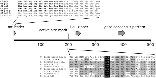
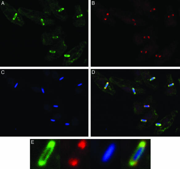
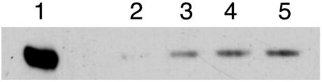
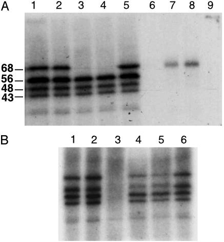
Kinetoplast DNA (kDNA), the form of mitochondrial DNA in trypanosomatids, consists of thousands of interlocked circular DNAs organized into a compact disk structure. A type II DNA topoisomerase, a DNA polymerase beta, and a structure-specific endonuclease have been localized to antipodal sites flanking the kDNA disk along with nascent DNA minicircles. We have cloned a gene (LIG k) encoding a mitochondrial DNA ligase in the trypanosomatid Crithidia fasciculata, and we show that an epitope-tagged form of the ligase colocalizes with the other replication proteins at the antipodal sites and also at the two faces of the kDNA disk. DNA LIG k becomes adenylated in reactions with ATP, and the adenylate moiety is removed by incubation with pyrophosphate or nicked DNA. The ligase interacts physically with the beta polymerase and is proposed to be involved in the repair of gaps in the newly synthesized minicircles. In yeast and mammals, a single gene encodes both nuclear and mitochondrial forms of DNA ligase. The LIG K protein sequence has low similarity to mitochondrial DNA ligases in other eukaryotes and is distinct from the C. fasciculata nuclear DNA ligase (LIG I).


Figures

Fig. 1.
Fig. 1.
Schematic representation of the 502-aa C. fasciculata mitochondrial DNA ligase protein and protein-sequence alignments. The N-terminal sequences of the C. fasciculata and T. brucei mitochondrial ligases are shown above the schematic diagram aligned with N-terminal sequences of C. fasciculata proteins known to have cleavable leader sequences (in bold). Sequences around the ligase active-site motif are shown below the schematic diagram aligned with those of several known mitochondrial (LIG III) and nuclear (LIG I) DNA ligases. The active-site lysine is indicated in white text on a black background. A ligase-consensus pattern (prosite) and a leucine-zipper sequence are indicated by arrows. GenBank accession nos. are as follows: AAB25701 (p16), AY143553 (p17), AF008943 (p18), AAC32801 (p21), AAA68599 (polymerase β), NM002311 (Homo sapiens LIG IIIα), MMU66058 (Mus musculus LIG IIIα), AF393654 (Xenopus laevis lig IIIα), CAA80615 (C. fasciculata LIG I), AAQ88427 (C. fasciculata LIG k), AC007863 (T. brucei LIG I), AAQ72485 (T. brucei LIG kα), AAA59518 (H. sapiens LIG I), and AAC75464 (Escherichia coli LIG).
Fig. 2.
Fig. 2.
Immunolocalization of the C. fasciculata DNA ligase. C. fasciculata cells transformed with pMS50.4 expressing the HA epitope-tagged DNA ligase were immunostained with mouse mAbs against the HA epitope and with rabbit polyclonal Abs against the mitochondrial DNA polymerase β.(A) Immunofluorescence of LIG k. (B) Immunofluorescence of polymerase β. (C) DNA fluorescent stain. (D) Merged images. (E) Higher magnification of fluorescence from a single kinetoplast.
Fig. 3.
Fig. 3.
Epitope-tagged mitochondrial DNA ligase coimmunoprecipitates with the mitochondrial DNA polymerase β. Lane 1 shows cell lysate corresponding to 1.5 × 10⁶ cells. Cell lysate corresponding to 3 × 10⁸ cells was immunoprecipitated with 0, 0.5, 1.0, and 2.0 μl of Abs against DNA polymerase β in lanes 2–5, respectively. The blot was probed with 12CA5 mAbs against the HA-epitope tag.
Fig. 4.
Fig. 4.
(A) Immunoprecipitation of adenylated DNA ligase. Lane 1 shows adenylation of phosphocellulose purified proteins. Lanes 2–5, show supernatants from immunoprecipitation reactions incubated with 0, 0.1, and 0.5 μlof 12CA5 mAbs or 1 μl of anti-CSBPB (44), respectively. Lanes 6–9 show immunoprecipitates from the reactions run in lanes 2–5. (B) Deadenylation of HA-tagged DNA ligase. The adenylated proteins (lane 1) were mock incubated (lane 2) or incubated with sodium pyrophosphate (lane 3), poly(dA)·oligo(dT) (lane 4), DNase I-treated calf thymus DNA (lane 5), or poly(rA)·oligo(dT) (lanes 6).

Comment in

  • Closing the gaps in kinetoplast DNA network replication.
    Klingbeil MM, Englund PT. Klingbeil MM, et al. Proc Natl Acad Sci U S A. 2004 Mar 30;101(13):4333-4. doi: 10.1073/pnas.0401400101. Epub 2004 Mar 22. Proc Natl Acad Sci U S A. 2004. PMID: 15070715 Free PMC article. Review. No abstract available.

References

    1. Shapiro, T. A. & Englund, P. T. (1995) in Annu. Rev. Microbiol., 49, 117–143. - PubMed
    1. Klingbeil, M. M., Drew, M. E., Liu, Y., Morris, J. C., Motyka, S. A., Saxowsky, T. T., Wang, Z. & Englund, P. T. (2001) Protist 152, 255–262. - PubMed
    1. Morris, J. C., Drew, M. E., Klingbeil, M. M., Motyka, S. A., Saxowsky, T. T., Wang, Z. & Englund, P. T. (2001) Int. J. Parasitol. 31, 453–458. - PubMed
    1. Sturm, N. R. & Simpson, L. (1990) Cell 61, 879–884. - PubMed
    1. Chen, J., Rauch, C. A., White, J. H., Englund, P. T. & Cozzarelli, N. R. (1995) Cell 80, 61–69. - PubMed

Publication types

Associated data

LinkOut - more resources