. 2004 Mar 30;101(13):4401-6.
doi: 10.1073/pnas.0400805101.

Functional association between Wwox tumor suppressor protein and p73, a p53 homolog

Affiliations

Functional association between Wwox tumor suppressor protein and p73, a p53 homolog

Rami I Aqeilan et al. Proc Natl Acad Sci U S A. .

Abstract

The WWOX gene is a recently cloned tumor suppressor gene that spans the FRA16D fragile region. Wwox protein contains two WW domains that are generally known to mediate protein-protein interaction. Here we show that Wwox physically interacts via its first WW domain with the p53 homolog, p73. The tyrosine kinase, Src, phosphorylates Wwox at tyrosine 33 in the first WW domain and enhances its binding to p73. Our results further demonstrate that Wwox expression triggers redistribution of nuclear p73 to the cytoplasm and, hence, suppresses its transcriptional activity. In addition, we show that cytoplasmic p73 contributes to the proapoptotic activity of Wwox. Our findings reveal a functional cross-talk between p73 and Wwox tumor suppressor protein.


Figures

Fig. 1.
Fig. 1.
Association of Wwox and p73. (A) 293 cells were transiently transfected with the expression plasmids encoding HA-p73α, HA-p73β, or HA-ΔNp73 (as indicated at the top) and Myc-Wwox. Thirty-six hours after transfection, whole-cell lysates were immunoprecipitated (IP) with anti-HA, anti-IgG, or anti-Myc antibodies. The immunoprecipitates were analyzed by immunoblotting (IB) with anti-HA (Upper) or anti-Myc (Lower) antibodies. (B) 293 cells were incubated with 50 μM etoposide for 16 h, and lysates were immunoprecipitated with anti-p73, anti-IgG, or monoclonal anti-wwox antibodies, followed by immunoblotting with anti-p73 (Upper) or anti-wwox (Lower). (C) p53 and Myc-Wwox were coexpressed in 293 cells and immunoprecipitated by anti-p53, anti-IgG, or anti-Myc antibodies, followed by immunoblotting with anti-p53 (Upper) or anti-Myc (Lower). (D and E) 293 cells were transiently transfected with the HA-p73β, HA-p73βY487A (D Lower), or HA-p73γ (E) and Myc-Wwox expression plasmids. Immunoprecipitation and immunoblotting were as in A. (F) MCF-7 cells were cotransfected with HA-p73β (Upper) or HA-p73γ (Lower). Lysates were immunoprecipitated with anti-HA, anti-IgG, or monoclonal anti-wwox antibodies, followed by immunoblotting with anti-HA. (G) 293 cells were transiently transfected with the different Wwox expression constructs (as indicated at the top) and HA-p73β. Immunoprecipitation and immunoblotting were as in A.(H) 293 cells were transfected with HA-p73β and lysates were mixed with the indicated GST fusions or GST alone. Complexes were captured with glutathione-Sepharose, and bound protein was detected by HA immunoblot. (I) 293 cells were transfected with HA-p73β or HA-p73γ, and lysates were mixed with GST-WI-WT or GST alone and then treated as in H.
Fig. 2.
Fig. 2.
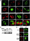
Subcellular localization of Wwox and p73. (A) NIH 3T3 cells were transfected as follows. (a) HA-p73β alone; (b) Myc-Wwox alone; (c1) Myc-Wwox; (c2) and HA-p73β; (d1) Myc-WWY33R; (d2) HA-p73β; (e1) Myc-WWY61R; (e2) HA-p73β. After fixation and permeabilization, the cells were stained with anti-HA and anti-Myc, followed by FITC-conjugated anti-mouse IgG (green; p73) and Texas red-conjugated anti-rabbit IgG (red; Wwox). (B) MCF-7 cells were transfected as follows. (f) empty vector; (g) HA-p73α; (h) HA-ΔNp73; (i) HA-p73γ; (j) HA-p73βY487A; (k) p53. Cells were prepared as in A. Antibodies used were polyclonal anti-wwox (f, g1, h1, i, j, and k), anti-HA (g2, h2, i, and j), and anti-p53 (k). (C and D) Effect of RNA interference directed against endogenous Wwox on p73 subcellular localization. MCF-7 cells were transfected with 100 nM siRNA as indicated. Forty-eight hours after treatment with siRNA, cells were transfected with HA-p73α plasmid for another 24 h. (C) Western blot analysis of siRNA effect on endogenous Wwox expression. (D) Fluorescence images showing Wwox-specific RNA interference effects in MCF-7 cells. Wwox is in red and p73 is in green. (Top) Images of mock-treated cells (no siRNA added). (Bottom) Images of irrelevant siRNA-treated cells. (Middle) Images of Wwox siRNA-treated cells.
Fig. 3.
Fig. 3.
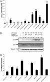
Wwox–p73 complexes alter p73 function. (A and B) Wwox suppresses p73-mediated transcriptional activity. (A) SAOS-2 cells were transiently cotransfected with the luciferase reporter construct (0.1 μg) carrying the p53/p73-responsive element derived from the p53 promoter and the indicated amounts of the following: bar 1, empty vector; bars 2 and 9, HA-p73β; bars 3 and 4, Myc-Wwox; bar 5, Myc-WWY33R; bars 6, 7, 10, and 11, HA-p73β and Myc-Wwox; bars 8 and 12, HA-p73β and Myc-WWY33R. Empty vector was cotransfected to normalize plasmid concentration where required. Twenty-four hours after transfection, cells were lysed and luciferase activity was determined. Results are shown as fold induction of the luciferase activity compared to control cells transfected with empty vector alone and are the average of three experiments. (B) Transfected SAOS-2 cells in A were assayed for Wwox and p73 protein expression by using anti-Myc and anti-HA antibodies, respectively. The same membrane was immunoblotted with anti-p21 and anti-tubulin antibodies. (C) Interaction between Wwox and p73 contributes to Wwox proapoptotic activity. SAOS-2 cells were transfected with the indicated combinations together with pEGFP vector (1:10) as control for transfection efficiency. After 48 h, cells were collected, fixed, and analyzed by fluorescence-activated cell sorting for DNA content versus cell number, using propidium iodide. Results are shown as average ± SD and are from three experiments. The results are expressed as the percentage of cells with sub-G1 DNA.
Fig. 4.
Fig. 4.
Phosphorylation of Wwox enhances its association with p73. (A) 293 cells were transfected with HA-p73β, Myc-Wwox, and Act-Src or DN-Src. Thirty-six hours later, whole-cell lysates were immunoprecipitated with anti-HA, anti-IgG, or anti-Myc. The immunoprecipitates were analyzed by immunoblotting with anti-HA. (B) Immunoprecipitates from A were immunoblotted with anti-HA (lanes1 and 2), anti-Myc (lanes 3 and 4), and anti-phosphotyrosine (lanes 5–8). (C) 293 cells were transfected with HA-p73β, Myc-Wwox, or Myc-WWY33R and Src as indicated. Immunoprecipitation and immunoblotting were performed as in A. (D) Lysates from C were immunoprecipitated with anti-Myc and immunoblotted with anti-phosphotyrosine (Upper) and anti-Myc (Lower). (E) Purified GST-polypeptide fusions with the different mutations were incubated with recombinant Src and ATP for 0 or 30 min. Reaction products were analyzed by SDS/PAGE and blotted with anti-phosphotyrosine.

References

    1. Bednarek, A. K., Laflin, K. J., Daniel, R. L., Liao, Q., Hawkins, K. A. & Aldaz, C. M. (2000) Cancer Res. 60, 2140–2145. - PubMed
    1. Paige, A. J., Taylor, K. J., Taylor, C., Hillier, S. G., Farrington, S., Scott, D., Porteous, D. J., Smyth, J. F., Gabra, H. & Watson, J. E. (2001) Proc. Natl. Acad. Sci. USA 98, 11417–11422. - PMC - PubMed
    1. Ried, K., Finnis, M., Hobson, L., Mangelsdorf, M., Dayan, S., Nancarrow, J. K., Woollatt, E., Kremmidiotis, G., Gardner, A., Venter, D., et al. (2000) Hum. Mol. Genet. 9, 1651–1663. - PubMed
    1. Kuroki, T., Trapasso, F., Shiraishi, T., Alder, H., Mimori, K., Mori, M. & Croce, C. M. (2002) Cancer Res. 62, 2258–2260. - PubMed
    1. Yendamuri, S., Kuroki, T., Trapasso, F., Henry, A. C., Dumon, K. R., Huebner, K., Williams, N. N., Kaiser, L. R. & Croce, C. M. (2003) Cancer Res. 63, 878–881. - PubMed

Publication types

MeSH terms