. 2004 Mar 30;101(13):4435-40.
doi: 10.1073/pnas.0306786101. Epub 2004 Feb 26.

Exploring amyloid formation by a de novo design

Affiliations

Exploring amyloid formation by a de novo design

Richard A Kammerer et al. Proc Natl Acad Sci U S A. .

Abstract

Protein deposition as amyloid fibrils underlies many debilitating human disorders. The complexity and size of disease-related polypeptides, however, often hinders a detailed rational approach to study effects that contribute to the process of amyloid formation. We report here a simplified peptide sequence successfully designed de novo to fold into a coiled-coil conformation under ambient conditions but to transform into amyloid fibrils at elevated temperatures. We have determined the crystal structure of the coiled-coil form and propose a detailed molecular model for the peptide in its fibrillar state. The relative stabilities of the two structural forms and the kinetics of their interconversion were found to be highly sensitive to small sequence changes. The results reveal the importance of specific packing interactions on the kinetics of amyloid formation and show the potential of this exceptionally favorable system for probing details of the molecular origins of amyloid disease.


Figures
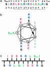
Fig. 1.
Fig. 1.
Design of the ccβ model system. (a) Amino acid sequence of ccβ. Positions X7 and X14 are occupied by Ala and Leu residues, respectively, in ccβ-p and by Met residues in ccβ-Met. The heptad repeats (abcdefg), capping residues (Ncap-acetyl and Ccap-amide), and the sequence patterning of polar (p) and hydrophobic (h) residues are indicated. (b) Helical wheel representation of ccβ as seen along the helix axis from the N terminus. (c) Schematic representation of ccβ in its β-strand conformation as seen along the β-sheet plane. Residues are colored according to their physicochemical properties: blue, positively charged; red, negatively charged; green, hydrophobic; black, polar and Gly.
Fig. 2.
Fig. 2.
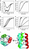
Structural and kinetic analysis of ccβ variants. (a) CD spectra recorded from ccβ-p (0.2 mg/ml) at 4°C (•) and after incubation for 3 days at 37°C (○). (b) Urea-induced unfolding profiles of 0.15 mg/ml ccβ-p (•), ccβ-Met (▴), and ccβ-MetO (▪) monitored by CD at 222 nm and 4°C. (c) A 2-Å resolution crystal structure of ccβ-p. Only the amino acid side chains at a, d, e, and g positions are shown. (d) α-to-β transition of ccβ-p (0.2 mg/ml) monitored at 37°CbyCD at 205 nm in the absence (•) and presence (○) of 5% (wt/wt) preformed fibrils. (e) α-to-β transition of ccβ derivatives and peptide mixtures monitored by OD at 350 nm and 37°C: 0.4 mg/ml ccβ-Met (•), 0.2 mg/ml ccβ-Met (○), equimolar mixture of ccβ-Met and ccβ-p, 0.2 mg/ml each (▴), and equimolar mixture of ccβ-Met and ccβ-MetO, 0.2 mg/ml each (▪). All spectroscopic data were obtained in PBS.
Fig. 3.
Fig. 3.
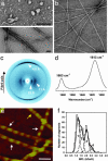
Structural analysis of ccβ amyloid aggregates. (a) TEM micrograph of negatively stained ccβ-p protofibrillar intermediates (Upper) and mature ccβ-p fibrils (Lower) obtained in PBS. (b) TEM micrograph of negatively stained mature ccβ-p fibrils obtained in water. (c) X-ray diffraction image of ccβ-p fibrils obtained with the beam perpendicular to the fiber axis. (d) Fourier transform infrared spectrum in the amide I region recorded from ccβ-p fibrils. (e) ccβ-Met fibrils imaged by AFM in aqueous solution. The arrows indicate the two prominent fibril populations, which are characterized by 590 ± 50 Å (white arrows) and 300 ± 50 Å (yellow arrow) periodicities. (f) STEM-MPL histogram of unstained and freeze-dried ccβ-Met fibrils. The data could be fitted well by four Gaussian curves peaking at 1.17 (peak 1), 1.55 (peak 2), 2.06 (peak 3), and 2.51 kDa/Å (peak 4) with an SD for each peak of 0.16 kDa/Å. (Scale bars, 500 Å.)
Fig. 4.
Fig. 4.
Proposed ccβ fibril model. View of four β-strands perpendicular to the plane of the β-sheets (a) and of the fibril cross section (b). Adjacent strands are related by a twofold screw axis along (a) and perpendicular to (b) the long fibril axis. Amino acid side chains are represented as spheres; the color code is the same as in Fig. 1. The black and gray arrows discriminate between the two faces of the β-strand. Residues at positions 7 and 14 are marked by white and yellow crosses, respectively. (c) REDOR difference signal ΔS/S0 from a sample of ccβ fibrils with 15N label at the amide nitrogen of Ala-7 and 13C label at the carbonyl carbon of Leu-14 as a function of dephasing time τREDOR. Experimental REDOR difference data (•) were obtained from the peak intensities of the carbonyl resonance. The SD for each data point was ± 0.01. The red curve was calculated by assuming an idealized antiparallel structure with extended β-strands and the register being defined by a hydrogen bond between the amide of Ala-7 and the carbonyl of Leu-14. The agreement of the simulation to the data is excellent for the important short dephasing times <20 ms where the transfer is determined primarily by the shorter distance dNC. The blue curve was calculated for a structure in which the register of the strands has been shifted by one residue. (d) REDOR measurements (•) on a 12% double-labeled fibrillized ccβ-p sample. The red and blue curves were calculated for extended interchain hydrogen-bonded β-strands and for a putative hairpin model with intrachain hydrogen bonds between the labeled residues, respectively.

Comment in

  • Unzipping the mysteries of amyloid fiber formation.
    Miranker AD. Miranker AD. Proc Natl Acad Sci U S A. 2004 Mar 30;101(13):4335-6. doi: 10.1073/pnas.0401163101. Epub 2004 Mar 22. Proc Natl Acad Sci U S A. 2004. PMID: 15070716 Free PMC article. Review. No abstract available.

References

    1. Taylor, J. P., Hardy, J. & Fischbeck, K. H. (2002) Science 296, 1991–1995. - PubMed
    1. Hardy, J. & Selkoe, D. J. (2002) Science 297, 353–356. - PubMed
    1. Guijarro, J. I., Sunde, M., Jones, J. A., Campbell, I. D. & Dobson, C. M. (1998) Proc. Natl. Acad. Sci. USA 95, 4224–4228. - PMC - PubMed
    1. Fändrich, M., Fletcher, M. A. & Dobson, C. M. (2001) Nature 410, 165–166. - PubMed
    1. Bucciantini, M., Giannoni, E., Chiti, F., Baroni, F., Formigli, L., Zurdo, J., Taddei, N., Ramponi, G., Dobson, C. M. & Stefani, M. (2002) Nature 416, 507–511. - PubMed

Publication types

Associated data

LinkOut - more resources