. 2004 Mar 30;101(13):4477-82.
doi: 10.1073/pnas.0306068101.

Hepatocyte growth factor/c-met signaling pathway is required for efficient liver regeneration and repair

Affiliations

Hepatocyte growth factor/c-met signaling pathway is required for efficient liver regeneration and repair

Chang-Goo Huh et al. Proc Natl Acad Sci U S A. .

Abstract

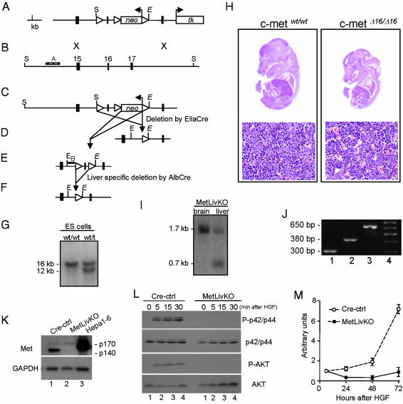
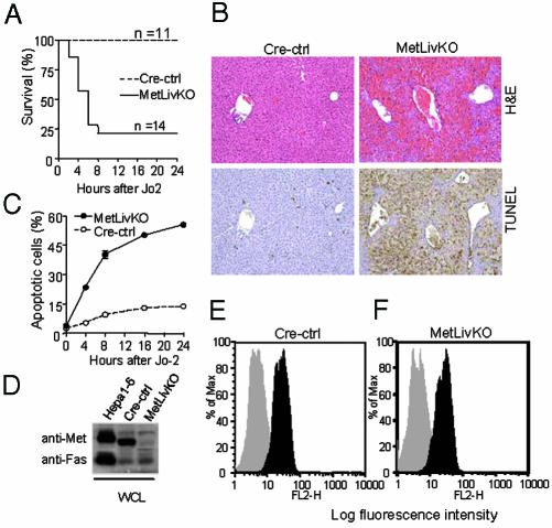
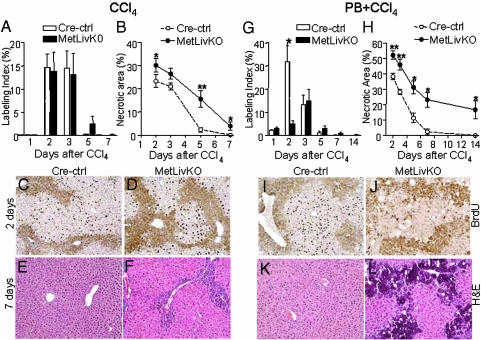
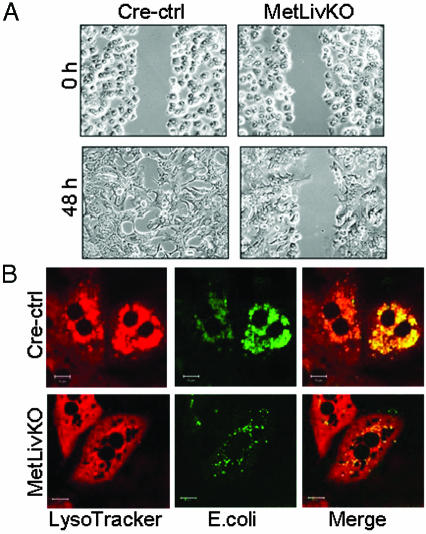
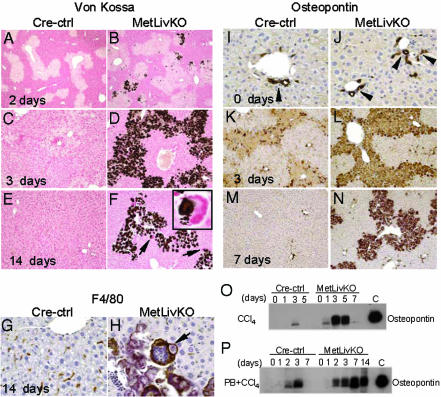
Hepatocyte growth factor/scatter factor c-met signaling pathway is of central importance during development as well as in tumorigenesis. Because homozygous null mice for either hgf/sf or c-met die in utero, we used Cre/loxP-mediated gene targeting to investigate the function of c-met specifically in the adult liver. Loss of c-met appeared not to be detrimental to hepatocyte function under physiological conditions. Nonetheless, the adaptive responses of the liver to injury were dramatically affected. Mice lacking c-met gene in hepatocytes were hypersensitive to Fas-induced apoptosis. When injected with a low dose of anti-Fas antibody, the majority of these mice died from massive apoptosis and hemorrhagic necrosis, whereas all wild-type mice survived with signs of minor injury. After a challenge with a single necrogenic dose of CCl4, c-met conditional knockout mice exhibited impaired recovery from centrolobular lesions rather than a deficit in hepatocyte proliferation. The delayed healing was associated with a persistent inflammatory reaction, over-production of osteopontin, early and prominent dystrophic calcification, and impaired hepatocyte scattering/migration into diseased areas. These studies provide direct genetic evidence in support of the critical role of c-met in efficient liver regeneration and suggest that disruption of c-met affects primarily hepatocyte survival and tissue remodeling.


Figures

Fig. 1.
Fig. 1.
Conditional deletion of exon 16 of mouse c-met gene. (A) c-met targeting vector. loxP sites are indicated by open triangles. neo, neomycin resistance gene; tk, herpes simplex thymidine kinase gene. (B) c-met wild-type allele. Filled boxes indicate c-met exons 15, 16, and 17. Hatched box indicates position of the probe A used for selection of targeted embryonic stem clones. (C) c-met targeted allele. S, SpeI; E, EcoRI. (D) Deleted allele (c-metΔ16) produced by crossing mice heterozygous for targeted allele (c-met+/t) to EllaCre transgenic mice. (E) Floxed allele (c-metfl). B, internal probe. (F) Liver-specific deletion of exon 16 by crossing c-metfl/fl mice to AlbCre transgenic mice. (G) Southern analysis of wild-type and targeted alleles. SpeI fragment specific to wild-type (16 kb) and targeted (12 kb) allele are shown. (H) c-met+/Δ16 and c-metΔ16/Δ16 embryos at embryonic day 14.5 and corresponding liver sections stained with hematoxylin/eosin. (Original magnification, ×25 and ×100, respectively.) (I) Southern analysis of tissue-specific deletion of floxed allele. The 1.7-kb floxed and 0.7-kb deleted alleles are shown. DNA extracted from 3-week-old MetLivKO mouse. (J) PCR analysis of genomic DNA from isolated hepatocytes. Lane 1, c-met+/+; lane 2, c-metfl/fl; lane 3, c-metfl/fl;AlbCre. PCR fragments corresponding to wild-type (300 bp), floxed (380 bp), and deletion-specific (650 bp) fragments are shown. (K) Western blot of cell lysates from cultured hepatocytes with indicated genotypes using anti-c-Met antibody. Hepa1–6, positive control. (L) Western blots showing phosphorylation status of p42/p44 MAPK and AKT upon HGF stimulation in cultured hepatocytes. (M) Response to rhHGF in primary hepatocyte cultures. Proliferation was measured in 96-well plates (1 × 10⁴ cells per well) by colorimetric assay (Roche Diagnostics).
Fig. 2.
Fig. 2.
The MetLivKo mice are more sensitive to Fas-mediated apoptosis. (A) Reduced survival in MetLivKO mice. (B) Hematoxylin and eosin and terminal deoxynucleotidyltransferase-mediated dUTP nick end labeling (TUNEL) staining 6 h after Jo-2 injection. (Original magnification, ×200.) (C) Assessment of apoptosis in vitro. Data represent cumulative average number of apoptotic cells ± SEM. At least 500 nuclei were counted from duplicate slides. (D) Western blot analysis of Met and Fas in whole cell lysates (WCL) from cultured hepatocytes. Hepa1–6, positive control. (E and F) Surface expression of Fas on freshly isolated hepatocytes determined by FACS (black histograms). Gray histograms, unstained cells.
Fig. 3.
Fig. 3.
c-met is required for liver healing but not hepatocyte proliferation. (A and G) Kinetics of BrdUrd incorporation. (B and H) Computer based morphometry of necrotic areas on hematoxylin/eosin-stained sections (NIH image). For each liver, necrotic areas within a 21-mm2 randomly selected field were measured. All data are means ± SEM; n = 5: *, P < 0.05; **, P < 0.001 as determined by Student's t test. (C, D, I, and J) BrdUrd immunostaining counterstained with heamotoxylin. (E, F, K, and L) Hematoxylin/eosin staining. (Original magnification, ×200.)
Fig. 4.
Fig. 4.
Lack of c-met signaling results in impairment of motility and phagocytosis. (A) Linear scrape wounds made in subconfluent monolayers of primary hepatocytes 4 h after plating were allowed to heal in the serum-free medium in the presence of 50 ng/ml rhHGF. (Original magnification, ×100.) (B) Representative confocal microscopy images of hepatocytes exposed to E. coli (green, Center) and lysosomal-specific fluorochrome (red, Left). Colocalization of green and red pixels appears as yellow (merge, Right). (Scale bar, 10 μm.)
Fig. 5.
Fig. 5.
Abnormalities of liver remodeling after injury with CCl4+PB. (AF) Calcium deposition was visualized by Von Kossa stain. (F Inset) Accumulation of calcium inside giant multinucleated cells shown by arrows. (G and H)F4/80 staining of Kupffer cells. Arrow points to a giant multinucleated Kupffer cell. (IN) Expression of osteopontin. (I and J) Osteopontin staining in biliary epithelial cells (arrowheads). Slides were counterstained with hematoxylin. (Original magnification, ×50 in A and B, ×100 in CF and KN, and ×400 in GJ and Inset in F). (O and P) Immunoblots with antiosteopontin. (P) Samples at time 0 were obtained after the last phenobarbital injection. C, recombinant mouse osteopontin (R & D Systems).

References

    1. Michalopoulos, G. K. & DeFrances, M. C. (1997) Science 276, 60–66. - PubMed
    1. Matsumoto, K. & Nakamura, T. (1996) J. Biochem. 119, 591–600. - PubMed
    1. Zhang, Y. W. & Vande Woude, G. F. (2003) J. Cell. Biochem. 88, 408–417. - PubMed
    1. Birchmeier, C. & Gherardi, E. (1998) Trends Cell Biol. 8, 404–410. - PubMed
    1. Vande Woude, G. F., Jeffers, M., Cortner, J., Alvord, G., Tsarfaty, I. & Resau, J. (1997) Ciba Found. Symp. 212, 119–130. - PubMed