. 2004 Mar 30;101(13):4560-5.
doi: 10.1073/pnas.0400983101. Epub 2004 Mar 19.

Human IL-23-producing type 1 macrophages promote but IL-10-producing type 2 macrophages subvert immunity to (myco)bacteria

Affiliations

Human IL-23-producing type 1 macrophages promote but IL-10-producing type 2 macrophages subvert immunity to (myco)bacteria

Frank A W Verreck et al. Proc Natl Acad Sci U S A. .

Abstract

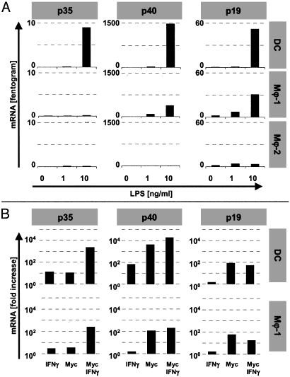
Macrophages (Mphi) play a central role as effector cells in immunity to intracellular pathogens such as Mycobacterium. Paradoxically, they also provide a habitat for intracellular bacterial survival. This paradoxical role of Mphi remains poorly understood. Here we report that this dual role may emanate from the functional plasticity of Mphi: Whereas Mphi-1 polarized in the presence of granulocyte-Mphi colony-stimulating factor promoted type 1 immunity, Mphi-2 polarized with Mphi colony-stimulating factor subverted type 1 immunity and thus may promote immune escape and chronic infection. Importantly, Mphi-1 secreted high levels of IL-23 (p40/p19) but no IL-12 (p40/p35) after (myco)bacterial activation. In contrast, activated Mphi-2 produced neither IL-23 nor IL-12 but predominantly secreted IL-10. Mphi-1 required IFN-gamma as a secondary signal to induce IL-12p35 gene transcription and IL-12 secretion. Activated dendritic cells produced both IL-12 and IL-23, but unlike Mphi-1 they slightly reduced their IL-23 secretion after addition of IFN-gamma. Binding, uptake, and outgrowth of a mycobacterial reporter strain was supported by both Mphi subsets, but more efficiently by Mphi-2 than Mphi-1. Whereas Mphi-1 efficiently stimulated type 1 helper cells, Mphi-2 only poorly supported type 1 helper function. Accordingly, activated Mphi-2 but not Mphi-1 down-modulated their antigen-presenting and costimulatory molecules (HLA-DR, CD86, and CD40). These findings indicate that (i) Mphi-1 and Mphi-2 play opposing roles in cellular immunity and (ii) IL-23 rather than IL-12 is the primary type 1 cytokine produced by activated proinflammatory Mphi-1. Mphi heterogeneity thus may be an important determinant of immunity and disease outcome in intracellular bacterial infection.


Figures

Fig. 1.
Fig. 1.
Differentiation of human blood monocytes into functionally distinct Mϕ subsets, Mϕ-1 and Mϕ-2. (A) In contrast to mo.DCs, Mϕ-1 and Mϕ-2 highly expressed CD14 but showed no or only weak expression of CD1a or CD83 (the latter after activation by LPS) as determined by flow cytometry. Cytokine secretion was measured up to 72 h after stimulation with M. tuberculosis sonicate (myc) (▪), LPS (•), or medium control (♦). (B) Whereas activated Mϕ-1, similar to DCs, secreted high levels of IL-12p40, Mϕ-2 failed to secrete IL-12p40. (C) IL-10, in contrast, was most predominant in Mϕ-2. Similar cytokine profiles were obtained with cells from at least 10 independent donors.
Fig. 2.
Fig. 2.
Type 1 cytokine secretion by monocyte-derived Mϕ and mo.DCs. The capacity of mo.DCs, Mϕ-1, and Mϕ-2 to secrete IL-12 (p40/p35) and IL-23 (p40/p19) proteins was determined 24 h after microbial stimulation in the absence (black bars) or presence (gray bars) of 500 units/ml IFN-γ. Mϕ-1 secreted IL-23 but failed to produce IL-12 after activation with mycobacterial sonicate (10 μg/ml) or LPS (10 ng/ml) unless IFN-γ was added. Also, DCs showed IFN-γ-enhanced IL-12 secretion, but unlike Mϕ-1 they yielded decreased rather than elevated IL-23 levels with IFN-γ. Mϕ-2 failed to produce IL-12 or IL-23. Depicted are average protein levels plus standard deviation (n = 3; n = 5 for IL-12 in DCs and Mϕ-1).
Fig. 3.
Fig. 3.
Type 1 cytokine mRNA levels in monocyte-derived Mϕ and mo.DCs. (A) Quantitative reverse transcription–PCR analyses 8 h after LPS stimulation of Mϕ-1 revealed that expression of IL-12-specific p35, but not p40 or p19 mRNA, required costimulation with IFN-γ (500 units/ml). DCs showed low but significant transcription of p35 in response to LPS and profound levels of p40 and p19 mRNA. Mϕ-2 failed to induce any of these cytokine transcripts. (B) Whereas stimulation with 10 μg/ml mycobacterial sonicate strongly induced the production of p40 and p19 mRNA, high-level transcription of p35 required costimulation with IFN-γ. (Note that the y axes are logarithmic in B!) Stimulation indexes were calculated from the average level of mRNA from two (DCs) to four (Mϕ-1) experiments. Activation-induced p19 transcription was inhibited by IFN-γ in both mo.DCs and Mϕ-1.
Fig. 4.
Fig. 4.
Binding, uptake, and outgrowth of mycobacteria by Mϕ-1 and Mϕ-2. (A) Binding (at 0°C) and/or uptake (at 37°C) of an M. bovis bacillus Calmette–Guérin-GFP reporter strain was determined by flow cytometry. After 20 min, more Mϕ-2 than Mϕ-1 had acquired fluorescent mycobacteria (representative of three independent experiments). (B) Accordingly, over a wide range of multiplicities of infection (MOI) from 20 to 2.5 mycobacteria per host cell, Mϕ-2 displayed enhanced outgrowth of M. bovis bacillus Calmette–Guérin-lux at day 6 compared with Mϕ-1. Depicted are averaged luminescence signals as cps [+ standard deviation (n = 4)]. Similar results were found with cells from different donors in four independent experiments.
Fig. 5.
Fig. 5.
Antigen-presenting capacity of Mϕ-1 versus Mϕ-2. To determine the antigen-presenting capacity of Mϕ-1 and Mϕ-2, cells were pretreated (black bars) or not (gray bars) for 24 h with a (myco)bacterial stimulus before incubation with antigen and antigen-specific Th1 cells. Mo.DCs were included for control purposes. (A) In contrast to Mϕ-1 and mo.DCs, Mϕ-2 relatively poorly supported proliferation of the M. leprae-specific Th1 clone R2F10 toward protein or peptide antigen, which was reduced further after activation of Mϕ-2 by M. tuberculosis sonicate (myc). (B) Similar results were obtained when antigen-presenting cells were pretreated with LPS. (C) The antigen-presenting capacity of Mϕ-2 toward the influenza hemagglutinin-specific T cell clone HA1.7 was reduced also by mycobacterial stimulation in a dose-dependent fashion. (D) Protein or peptide antigen-specific IFN-γ secretion of R2F10 Th1 cells was supported by Mϕ-1, albeit less efficiently than by mo.DCs. IFN-γ secretion by R2F10 cells responding to Mϕ-2 was substantially lower and reduced further by mycobacterial activation of Mϕ-2. IFN-γ production is depicted as secreted protein in the pooled supernatant of triplicate cultures. Proliferation is depicted as the average incorporation of [3H]thymidine in triplicate cultures (cpm; + standard deviation). Experiments were repeated at least twice using independent donors to generate Mϕ and DCs.
Fig. 6.
Fig. 6.
Expression of HLA and costimulatory molecules on Mϕ-1 and Mϕ-2. Mϕ were pretreated (gray bars) or not (black bars) with mycobacterial sonicate for 48 h before flow-cytometric analysis of HLA-DR, CD80, and CD86 expression. Mϕ-2 but not Mϕ-1 or mo.DCs down-regulated their cell surface levels of HLA-DR and the costimulatory molecule CD86 (but not CD80) after activation. Depicted are the mean fluorescent intensities (mfi; + standard deviation) of cells from three independent donors.

References

    1. Ottenhoff, T. H., Verreck, F. A., Lichtenauer-Kaligis, E. G., Hoeve, M. A., Sanal, O. & Van Dissel, J. T. (2002) Nat. Genet. 32, 97–105. - PubMed
    1. Kaufmann, S. H. (2001) Nat. Rev. Immunol. 1, 20–30. - PubMed
    1. Janeway, C. A., Jr., & Medzhitov, R. (2002) Annu. Rev. Immunol. 20, 197–216. - PubMed
    1. Sieling, P. A. & Modlin, R. L. (2002) Curr. Opin. Microbiol. 5, 70–75. - PubMed
    1. Trinchieri, G. (1995) Annu. Rev. Immunol. 13, 251–276. - PubMed

Publication types