. 2004 Mar 30;101(13):4608-13.
doi: 10.1073/pnas.0306928101. Epub 2004 Mar 18.

Mycobacterial polyketide-associated proteins are acyltransferases: proof of principle with Mycobacterium tuberculosis PapA5

Affiliations

Mycobacterial polyketide-associated proteins are acyltransferases: proof of principle with Mycobacterium tuberculosis PapA5

Kenolisa C Onwueme et al. Proc Natl Acad Sci U S A. .

Erratum in

  • Proc Natl Acad Sci U S A. 2004 Apr 27;101(17):6834

Abstract

Mycobacterium tuberculosis (Mt) produces complex virulence-enhancing lipids with scaffolds consisting of phthiocerol and phthiodiolone dimycocerosate esters (PDIMs). Sequence analysis suggested that PapA5, a so-called polyketide-associated protein (Pap) encoded in the PDIM synthesis gene cluster, as well as PapA5 homologs found in Mt and other species, are a subfamily of acyltransferases. Studies with recombinant protein confirmed that PapA5 is an acyltransferase [corrected]. Deletion analysis in Mt demonstrated that papA5 is required for PDIM synthesis. We propose that PapA5 catalyzes diesterification of phthiocerol and phthiodiolone with mycocerosate. These studies present the functional characterization of a Pap and permit inferences regarding roles of other Paps in the synthesis of complex lipids, including the antibiotic rifamycin.


Figures

Fig. 1.
Fig. 1.
Conservation of Hx3Dx14Y motif. Protein names are based on similarity to Mt Paps, except for Rif20 (with highest similarity to PapA5). Numbers in front of each protein segment indicate position of the conserved His. Total number of residues of each protein is indicated. Percent similarity of full-length Paps with their closest Mt Pap homolog is shown. Sequence alignment and similarity values were obtained with Clustal method (49). Am, A. mediterranei S699; Ma, Mycobacterium avium; Mp, M. avium subsp. paratuberculosis; Mb, M. bovis; Ml, M. leprae; Mm, Mycobacterium marinum; Ms, Mycobacterium smegmatis; Mt, M. tuberculosis; and Sc, S. coelicolor A3 (2). Two additional Paps from Mm are not shown.
Fig. 2.
Fig. 2.
Purified PapA5 and PapA5 variants. Coomassie blue-stained SDS/PAGE (12%) of purified PapA5 (lane 2), PapH123A (lane 3), PapH124A (lane 4), PapD128A (lane 5), and PapY143F (lane 6) and molecular marker (lane 1).
Fig. 3.
Fig. 3.
AT assays revealing PapA5 activity. (A) TLC analysis of AT reactions. Reactions with 18 μM[14C]PCoA, 180 μM 1-OCL, 75 mM Mes (pH 6.5) 100 mM NaCl, and PapA5 (1), no protein (2), BSA (3), His-6-tagged P. aeruginosa arylation enzyme PchD (4), His-6-tagged Mt synthetase MbtB ArCP domain (5), UlpI (6), or CAT (7) were incubated for 8 h. Analysis of reactions with PapA5 and [14C]PCoA but lacking 1-OCL (8), PapA5 and [14C]hexadecanol but lacking PCoA (9) and PapA5 [14C]PCoA and hexadecanol (10) is shown. Products octyl-palmitate (OP) in section 1, hexadecyl-palmitate (HP) in section 10, palmitate (PA) in all sections except 9, where the spot is unreacted [14C]hexadecanol, and TLC origin (Or) are marked. Assays were done in triplicate. Only duplicates are shown in sections 8-10. (B) ESI-MS of low Rf product from section 1 showing PA mass (m/z 256-H). (C) ESI-MS of high Rf products from section 1 showing OP mass (m/z 368 + Na).
Fig. 4.
Fig. 4.
PapA5-catalyzed octyl-palmitate formation is time- and pH-dependent. (A) Time course of product formation. (B) pH effect on product formation. Formation of octyl-palmitate (solid circles) and palmitate (open circles) is expressed as percentage of [14C]PCoA initial concentration. Reactions contained 2 μM PapA5, 18 μM[14C]PCoA, 180 μM 1-OCL, 100 mM NaCl, and 75 mM buffer (Mes, pH 6.5, for the time course and Mes, pH 6.0 or 6.5, Hepes, pH 7.0, and Tris·HCl, pH 7.5, 8.0, or 8.5, for pH-dependence analysis). Reactions were incubated for 12 h for pH-dependence analysis. Average of duplicates ± SE is shown.
Fig. 5.
Fig. 5.
Effect of 1-OCL and PCoA concentration on the rate of PapA5-catalyzed octyl-palmitate formation. (A) Reaction velocity as a function of 1-OCL concentration with [14C]PCoA at 18 μM. (B) Reaction velocity as a function of [14C]PCoA concentration with 1-OCL at 1 mM. Reactions contained substrates, 0.2 μM PapA5, 100 mM NaCl, and 75 mM Mes, pH 6.5, and were incubated for 3 h. Means of triplicates ± SEM are shown.
Fig. 6.
Fig. 6.
Activity of PapA5 mutant variants. TLC analysis showing ester formed in reactions with 18 μM[14C]PCoA/1 mM 1-OCL/75 mM Mes, pH 6.5/100 mM NaCl/0.2 μM enzyme incubated 3 h (Upper) or 2 μM enzyme incubated 24 h (Lower). Picomoles of ester are shown as mean of triplicates ± SEM. ND, not detected.
Fig. 7.
Fig. 7.
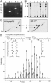
Deletion of papA5 and lipid profiling. (A) Confirmation of papA5 deletion. Lanes 1 and 6, molecular marker. Lanes 2 and 3, PCR products with primers 5xpf and 5xpr from genomic DNA of WT and ΔpapA5 Mt, respectively. Lanes 4 and 5, PCR products with primers 5ufx and 5urx from genomic DNA of WT and ΔpapA5 Mt, respectively. (B) 1D TLC of 14C-labeled apolar lipids of WT Mt (lane 1), ΔpapA5 (lane 2), ΔpapA5 with pMV261 (lane 3), ΔpapA5 with pCPapA5 (lane 4), ΔpapA5 with pCPH124 (lane 5), and ΔpapA5 with pCPY143 (lane 6). Spots I and II are POL- and PONE-dimycocerosate, respectively. TLC origin (Or) is marked. (C) 2D TLC of 14C-labeled apolar lipids of WT and ΔpapA5 Mt. PDIM spots and origin are labeled as in B. (D) Atmospheric pressure photoionization MS of total PDIMs corresponding to TLC spots missing in ΔpapA5 Mt. POL monoester ion (C63H125O3+) expected mass: 929.97 ± 14(n+m). PONE monoester ion (C62H121O3+) expected mass: 913.94 ± 14(n+m). (Inset) Ionization of POL (R, -OCH3) and PONE (R, =O) esters. l and m = 1-7, n = 0-2.

References

    1. Bentley, R. & Bennett, J. W. (1999) Annu. Rev. Microbiol. 53, 411-446. - PubMed
    1. Camacho, L. R., Constant, P., Raynaud, C., Laneelle, M. A., Triccas, J. A., Gicquel, B., Daffe, M. & Guilhot, C. (2001) J. Biol. Chem. 276, 19845-19854. - PubMed
    1. Cox, J. S., Chen, B., McNeil, M. & Jacobs, W. R. (1999) Nature 402, 79-83. - PubMed
    1. Rambukkana, A., Zanazzi, G., Tapinos, N., Salzer, J. L. (2002) Science 296, 927-931. - PubMed
    1. Cole, S. T., Brosch, R., Parkhill, J., Garnier, T., Churcher, C., Harris, D., Gordon, S. V., Eiglmeier, K., Gas, S., Barry, C. E., et al. (1998) Nature 393, 537-544. - PubMed

Publication types

MeSH terms

LinkOut - more resources