. 2004 Mar 30;101(13):4614-9.
doi: 10.1073/pnas.0400932101.

Salmonella typhi encodes a functional cytolethal distending toxin that is delivered into host cells by a bacterial-internalization pathway

Affiliations

Salmonella typhi encodes a functional cytolethal distending toxin that is delivered into host cells by a bacterial-internalization pathway

Erik Haghjoo et al. Proc Natl Acad Sci U S A. .

Abstract

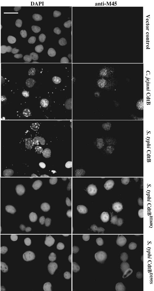
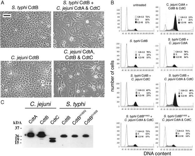
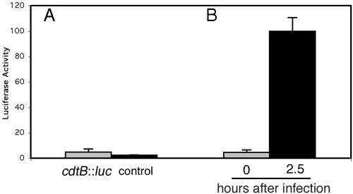
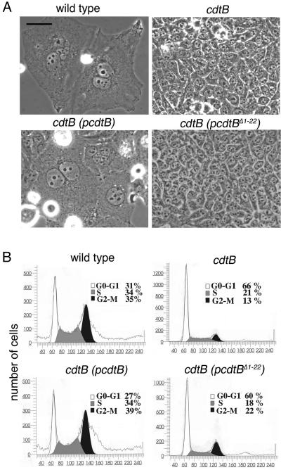
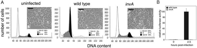
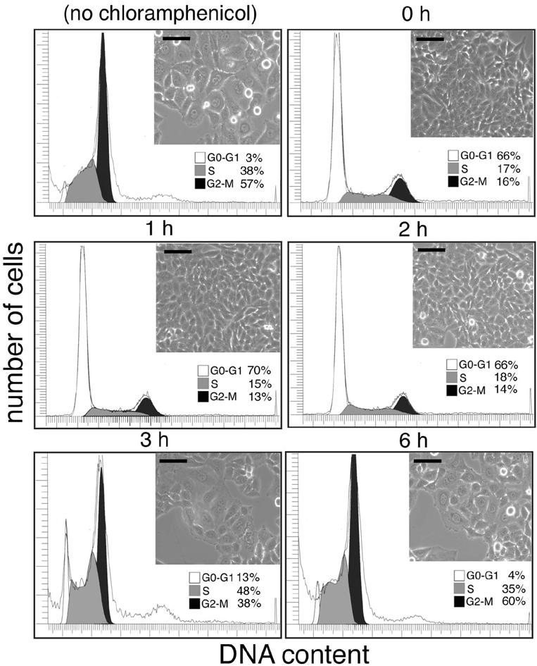
Many bacterial pathogens encode the cytolethal distending toxin (CDT), which causes host cells to arrest during their cell cycle by inflicting DNA damage. CDT is composed of three proteins, CdtA, CdtB, and CdtC. CdtB is the enzymatically active or A subunit, which possesses DNase I-like activity, whereas CdtA and CdtC function as heteromeric B subunits that mediate the delivery of CdtB into host cells. We show here that Salmonella enterica serovar Typhi encodes CDT activity, which depends on the function of a CdtB homologous protein. Remarkably, S. enterica serovar Typhi does not encode apparent homologs of CdtA or CdtC. Instead, we found that toxicity, as well as cdtB expression, requires bacterial internalization into host cells. We propose a pathway of toxin delivery in which bacterial internalization relieves the requirement for the functional equivalent of the B subunit of the CDT toxin.


Figures

Fig. 1.
Fig. 1.
Effect of transient expression of wild-type and catalytic mutants of S. typhi CdtB in cultured cells. Cos-2 cells were transfected with vectors encoding M45 epitope-tagged S. typhi CdtB, the catalytic mutants CdtBH160Q and CdtBD195S, or C. jejuni CdtB. Cells were stained 48 h after transfection with a monoclonal antibody directed to the M45 epitope tag (anti-M45) to visualize cells expressing the different CdtB proteins and with 4′,6-diamidino-2-phenylindole (DAPI) to visualize the chromatin. Images were obtained with a Nikon Eclipse inverted microscope fitted with a Princeton Instruments (Trenton, NJ) MicroMAX digital camera. (Scale bar, 50 μm.)
Fig. 2.
Fig. 2.
Effect of Cdt proteins on cultured intestinal epithelial cells. Extracts of E. coli strains (100 μg/ml) expressing different M45 epitope-tagged S. typhi or C. jejuni Cdt proteins as indicated, alone or in combination, were added to cultured Henle-407 cells. (A and B) Cells were examined 72 h after addition of the protein extracts under a phase microscope (A) or processed to measure DNA content by flow cytometry (B) as described in Materials and Methods. The peaks corresponding to cells in G0/G1 (G0-G1), S, or G2 (G2-M) are indicated. (C) The presence of the different Cdt proteins in the bacterial extracts was verified by Western blot analysis with an antibody directed to the M45 epitope tag. (Scale bar, 50 μm.)
Fig. 3.
Fig. 3.
Expression of S. typhi cdtB after growth in vitro or after infection of cultured cells. (A) A S. typhi strain (control) and an isogenic derivative encoding a cdtB::luc reporter fusion were grown in LB broth to an OD600 of 1.0, and the luciferase activity in total cell lysates was measured as described in Materials and Methods. (B) Alternatively, cultured intestinal Henle-407 cells were infected with the S. typhi cdtB::luc reporter strain, and the luciferase activity at different times after infection was measured as indicated in Materials and Methods. In all cases, values were standardized relative to the colony-forming units and the values obtained in cells 2.5 h after infection, which was considered 100%.
Fig. 4.
Fig. 4.
Effect of S. typhi infection of cultured intestinal epithelial cells. Henle-407 cells were infected with S. typhi, its isogenic cdtB mutant, or the cdtB mutant carrying a plasmid encoding either wild-type CdtB or a mutant lacking the putative sec-dependent secretion signal (pcdtBΔ1-22). Cells were examined 72 h after transfection under a phase microscope (A) or processed to measure DNA content by flow cytometry (B) as indicated in Materials and Methods. The peaks corresponding to cells in G0/G1 (G0-G1), S, or G2 (G2-M) are indicated. (Scale bar, 50 μm.)
Fig. 5.
Fig. 5.
Display of S. typhi CDT activity and expression of cdtB require bacterial internalization. Henle-407 cells were infected with S. typhi or its isogenic invA mutant, which is unable to enter host cells. (A) Cells were examined 72 h after infection under a phase microscope for signs of intoxication. (Scale bar, 50 μm.) In addition, cells were infected with derivatives of S. typhi or its isogenic invA mutant encoding the cdtB::luc reporter fusion strain, and the luciferase activity at different times after infection was measured as indicated in Materials and Methods. (B) In all cases, values were standardized relative to the colony-forming units and the values obtained in cells 2.5 h after infection, which was considered 100%.
Fig. 6.
Fig. 6.
Effect of bacterial protein synthesis inhibition on the display of CDT activity by S. typhi. Cultured intestinal Henle-407 cells were infected with the S. typhi strain, and the bacterial protein synthesis inhibitor chloramphenicol was added at different times after infection as indicated. Cells were examined 72 h after infection under a phase microscope for signs of intoxication or processed to measure DNA content by flow cytometry as indicated in Materials and Methods. The peaks corresponding to cells in G0/G1 (G0-G1), S, or G2 (G2-M) are indicated. (Scale bar, 50 μm.)

References

    1. Johnson, W. M. & Lior, H. (1988) Microb. Pathog. 4, 103-113. - PubMed
    1. Johnson, W. M. & Lior, H. (1988) Microb. Pathog. 4, 115-126. - PubMed
    1. Peres, S., Marches, O., Daigle, F., Nougayrede, J., Herault, F., Tasca, C., De Rycke, J. & Oswald, E. (1997) Mol. Microbiol. 24, 1095-1107. - PubMed
    1. Okuda, J., Kurazono, H. & Takeda, Y. (1995) Microb. Pathog. 18, 167-172. - PubMed
    1. Cope, L., Lumbley, S., Latimer, J., Klesney-Tait, J., Stevens, M., Johnson, L., Purven, M., Munson, R. J., Lagergard, T., Radolf, J. & Hansen, E. (1997) Proc. Natl. Acad. Sci. USA 94, 4056-4061. - PMC - PubMed

Publication types