. 2004 Mar 30;101(13):4706-11.
doi: 10.1073/pnas.0305574101. Epub 2004 Mar 22.

WIN1, a transcriptional activator of epidermal wax accumulation in Arabidopsis

Affiliations

WIN1, a transcriptional activator of epidermal wax accumulation in Arabidopsis

Pierre Broun et al. Proc Natl Acad Sci U S A. .

Abstract

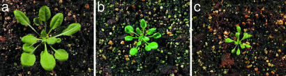
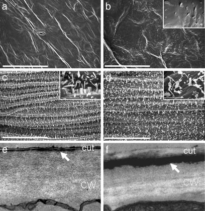
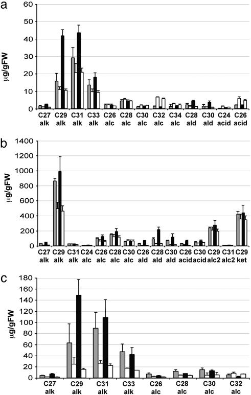
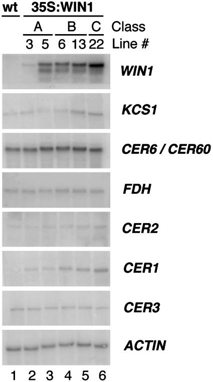
Epicuticular wax forms a layer of hydrophobic material on plant aerial organs, which constitutes a protective barrier between the plant and its environment. We report here the identification of WIN1, an Arabidopsis thaliana ethylene response factor-type transcription factor, which can activate wax deposition in overexpressing plants. We constitutively expressed WIN1 in transgenic Arabidopsis plants, and found that leaf epidermal wax accumulation was up to 4.5-fold higher in these plants than in control plants. A significant increase was also found in stems. Interestingly, approximately 50% of the additional wax could only be released by complete lipid extractions, suggesting that not all of the wax is superficial. Gene expression analysis indicated that a number of genes, such as CER1, KCS1, and CER2, which are known to be involved in wax biosynthesis, were induced in WIN1 overexpressors. This observation indicates that induction of wax accumulation in transgenic plants is probably mediated through an increase in the expression of genes encoding enzymes of the wax biosynthesis pathway.


Figures

Fig. 1.
Fig. 1.
Phenotype of WIN1 overexpressors. (a) Arabidopsis wild-type plant (Ecotype Columbia). (b) Class B 35S:WIN1 plant (line 6). (c) Class C plant (line 22). Arabidopsis plants overexpressing WIN1 show glossier leaves. Pictures were taken at the same magnification.
Fig. 2.
Fig. 2.
Electron microscopy analysis of 35S:WIN1 plants (line 22). (a and b) SEM images of leaves of wild-type (a) and 35S:WIN1 (b) plants. (b Inset) Plate-like wax crystals are observed in the 35S:WIN1 line. (c and d) SEM images of stems of control (c) and 35S:WIN1 (d) plants. (White bars in a-d, main image, 50 μm; white bars in Insets, 5 μm.) (c and d) TEM images of wild-type (e) and 35S:WIN1 (f) leaf epidermal cell sections. A thick layer of osmium dense material is observable in 35S:WIN1 epidermal cells underneath the epicuticular and peripheral cuticular layer (arrow). CW, cell wall; cut, cuticle. Images in e and f were taken at ×21,000 magnification.
Fig. 3.
Fig. 3.
Wax profile of selected tissues from transgenic 35S:WIN1 plants. (a) Leaf epicuticular wax composition. (b) Stem epicuticular wax composition. Leaf and stem wax constituents were extracted into chloroform, were TMS-derivatized, and were analyzed by GC-MS. (c) Quantitative analysis of total leaf wax. Wax components from methanolized leaf lipid extracts were analyzed by GC-MS after TMS derivatization. Each value for a wax constituent from a 35S:WIN1 line is followed by the corresponding value from control plants grown under the same conditions. All values are relative to fresh weight and represent the average of three independent experiments. Gray bars, line 6; black bars, line 22; and white bars, control line.
Fig. 4.
Fig. 4.
Expression of genes encoding wax biosynthetic enzymes in 35S:WIN1-transgenic lines. Northern analysis of 35S:WIN1 lines 3 and 5 (both class A), 6 and 13 (class B), and 22 (class C). Probes detected WIN1, KCS1, CER6/CUT1, and CER60 (two genes highly related in sequence, which could not be distinguished), FDH, CER2, CER1, and CER3; actin was used as control. Higher levels of CER1 and KCS1 transcripts are detected in the transgenic lines.
Fig. 5.
Fig. 5.
Analysis of WIN1 tissue expression. WIN1 expression was determined by semiquantitative RT-PCR in a variety of tissues from wild-type Columbia plants (Upper). RT-PCR was also performed with actin primers as control (Lower). WIN1 RT-PCR products are shown after 36 PCR cycles, and actin products are shown after 28 PCR cycles.

References

    1. Riederer, M. & Schreiber, L. (2001) J. Exp. Bot. 52, 2023-2032. - PubMed
    1. Kerstiens, G. (1996) J. Exp. Bot. 47, 1813-1832.
    1. Eigenbrode, S. D. & Espelie, K. E. (1995) Annu. Rev. Entomol. 40, 171-194.
    1. Belding, R. D., Sutton, T. B., Blankenship, S. M. & Young, E. (2000) Plant Dis. 84, 767-772. - PubMed
    1. Marcell, L. M. & Beattie, G. A. (2002) Mol. Plant-Microbe Interact. 15, 1236-1244. - PubMed

Publication types

MeSH terms

Associated data