. 2004 Apr;42(4):1570-6.
doi: 10.1128/JCM.42.4.1570-1576.2004.

Immunological characterization of the spike protein of the severe acute respiratory syndrome coronavirus

Affiliations

Immunological characterization of the spike protein of the severe acute respiratory syndrome coronavirus

Liqun Lu et al. J Clin Microbiol. 2004 Apr.

Abstract

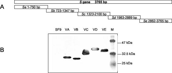
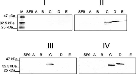
Severe acute respiratory syndrome (SARS) is a novel infectious disease caused by the SARS-associated coronavirus (SARS-CoV). There are four major structural proteins in the SARS-CoV, including the nucleocapsid, spike, membrane, and small envelope proteins. In this study, two sets of truncated fragments of spike protein were generated, the first were approximately 210-bp nonoverlapping fragments and the second were overlapping segments of 750 to 900 bp. From these 23 fragments, we identified a fragment of 259 amino acids (amino acids 441 to 700) that is a major immunodominant epitope. This fragment was highly expressed, and the purified fragment C could detect all 33 SARS patient serum samples tested, collected from 7 to 60 days after the onset of fever, but had no reactivity with all 66 healthy human serum samples tested. Thus, fragment C of spike protein was identified as an immunodominant antigen and could be used for serological detection of SARS-CoV infection.


Figures

FIG. 1.
FIG. 1.
SDS-PAGE and Western blot analysis of 18 nonoverlapping fragments covering the whole S protein expressed as GST fusion proteins. (A) Schematic diagram of 18 nonoverlapping fragments within the S gene. (B) SDS-PAGE of the 18 induced GST fusion fragments. GST fusion proteins around 32 kDa were expressed in all of the induced cells. M, protein marker; BL21, control BL21 cellular extract; S1 to S18, total cellular extracts harvested 4 h post-IPTG induction of the BL21 cells transformed with recombinant pGEX constructs bearing s1 to s18, respectively. (C) Western blot analysis of the expressed GST fusion fragments with monoclonal mouse anti-GST antibody. M, protein marker; GST, control GST protein; N, control BL21 cellular extract; S1 to S18, samples as described for panel A. The arrows indicate the expressed fusion proteins.
FIG. 2.
FIG. 2.
Expression of five overlapping domains within the S protein in SF9 cells. (A) Schematic diagram of five overlapping fragments within the S protein. (B) Western blot analysis of the expressed His6-tagged fragments. SF9, control SF9 cells; VA, fragment A; VB, fragment B; VC, fragment C; VD, fragment D; VE, fragment E; M, protein marker.
FIG. 3.
FIG. 3.
Representative immunoblot assays with sera from the 10 randomly selected SARS patients. Total cellular extracts of fragments A to E were subjected to SDS-12% PAGE and immunoblot analysis. For all of the immunoblot assays, sera were used at 1:100 and peroxidase-conjugated goat anti-total Ig polyclonal sera were used at 1:1,000. M, protein marker; SF9, cellular extracts from null-infected SF9 cells; A to E, cellular extracts from SF9 cells infected with VA, VB, VC, VD, and VE, respectively. (I) Immunoblot with one healthy human serum sample as a control. (II) Immunoblot with serum sample no. 1 that reacted with domains C, D, and E. (III) Immunoblot with serum sample no. 5 that recognized only the C domain of S protein. (IV) Immunoblot with serum sample no. 8 that recognized both the C and D domains.
FIG. 4.
FIG. 4.
Expression, purification, and immunoblot analysis of the His6-tagged C domain from E. coli strain M15. (A) SDS-PAGE of samples eluted from affinity chromatography purification. M, protein marker; lane 1, total cellular extract before IPTG induction; lane 2, total cellular extract 4 h post-IPTG induction; lane 3, eluted protein sample from affinity chromatography purification. (B) Immunoblot of fragment C with the monoclonal anti-His5 antibody. Lane 4, purified protein sample. (C) Immunoblot of purified fragment C with a pooled serum sample from the 10 positive serum samples. Lane 5, purified protein sample as described for lane 3.

References

    1. Callebaut, P., L. Enjuanes, and M. Pensaert. 1996. An adenovirus recombinant expressing the spike glycoprotein of porcine respiratory coronavirus is immunogenic in swine. J. Gen. Virol. 77:309-313. - PubMed
    1. Drosten, C., S. Gunther, W. Preiser, S. van der Werf, H. R. Brodt, S. Becker, H. Rabenau, M. Panning, L. Kolesnikova, R. A. Fouchier, A. Berger, A. M. Burguiere, J. Cinatl, M. Eickmann, N. Escriou, K. Grywna, S. Kramme, J. C. Manuguerra, S. Muller, V. Rickerts, M. Sturmer, S. Vieth, H. D. Klenk, A. D. Osterhaus, H. Schmitz, and H. W. Doerr. 2003. Identification of a novel coronavirus in patients with severe acute respiratory syndrome. N. Engl. J. Med. 348:1967-1976. - PubMed
    1. Enjuanes, L., et al. 2000. Coronaviridae, p. 845-849. In M. H. V. van Regenmortal, C. M. Fauqet, D. H. L. Bishop, E. B. Carstens, M. K. Estes, S. M. Lemon, M. A. Mayo, D. J. McGeoch, C. R. Pringle, and R. B. Wickner (ed.), Virus taxonomy. Academic Press, New York, N.Y.
    1. Fouchier, R. A., T. Kuiken, M. Schutten, G. van Amerongen, G. J. van Doornum, B. G. van den Hoogen, M. Peiris, W. Lim, K. Stohr, and A. D. Osterhaus. 2003. Aetiology: Koch's postulates fulfilled for SARS virus. Nature 423:240. - PMC - PubMed
    1. Gallagher, T. M., and M. J. Buchmeier. 2001. Coronavirus spike proteins in viral entry and pathogenesis. Virology 279:371-374. - PMC - PubMed

MeSH terms

LinkOut - more resources