. 2004 Apr 7;24(14):3511-21.
doi: 10.1523/JNEUROSCI.0290-04.2004.

Dendritic control of spontaneous bursting in cerebellar Purkinje cells

Affiliations

Dendritic control of spontaneous bursting in cerebellar Purkinje cells

Mary D Womack et al. J Neurosci. .

Abstract

We investigated the mechanisms that contribute to spontaneous regular bursting in adult Purkinje neurons in acutely prepared cerebellar slices. Bursts consisted of 3-20 spikes and showed a stereotypic waveform. Each burst developed with an increase in firing rate and was terminated by a more rapid increase in firing rate and a decrease in spike height. Whole-cell current-clamp recordings showed that each burst ended with a rapid depolarization followed by a hyperpolarization. Dual dendritic and somatic extracellular recordings revealed that each burst was terminated by a dendritic calcium spike. The contributions of T- and P/Q-type calcium current, large (BK) and small (SK) conductance calcium-activated potassium currents, and hyperpolarization-activated (I(H)) current to bursting were investigated with specific channel blockers. None of the currents, except for P/Q, were required to sustain spontaneous bursting or the stereotypic burst waveform. T-type calcium, BK, and SK channels contributed to interspike and interburst intervals. The effect of T-type calcium channel block was more pronounced after BK channel block and vice versa, indicating that these two currents interact to regulate burst firing. Block of I(H) current had no effect on bursting. Partial block of P/Q-type calcium channels concurrently eliminated dendritic calcium spikes and caused a switch from regular bursting to tonic firing or irregular bursting. Dendritic calcium spikes persisted in the presence of tetrodotoxin, indicating that their initiation did not require somatic sodium spikes. Our results demonstrate an important role for dendritic conductances in burst firing in intact Purkinje neurons.


Figures
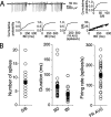
Figure 1.
Figure 1.
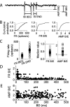
Characterization of spontaneous bursts in Purkinje neurons. A, Extracellular recording from a Purkinje neuron during spontaneous bursting. By analyzing bursts from several cycles of the trimodal pattern of activity, cumulative probability plots of spikes per burst (S/B), burst duration (BD), interburst interval (IBI), and average firing rate (FR AVG) were generated. Also shown is a plot of BD versus S/B. B, The median value of each parameter measured is shown for each cell (open circles). The mean of all the medians for each parameter is also shown (filled squares). Error bars represent ±SEM (n = 27).
Figure 2.
Figure 2.
Spontaneous bursts end with an increase in firing rate and a decrease in spike amplitude. Each burst was analyzed for changes in firing rate and spike amplitude. A, The diagram shows burst parameters that were measured. ISI BEG, First interspike interval; ISI END, last interspike interval; AMP BEG, first spike amplitude; AMP END, last spike amplitude. The instantaneous firing rate at the beginning (FR BEG) and end (FR END) of each burst was calculated by taking the reciprocal of the corresponding interspike interval. B, Cumulative probability plots of FR BEG (thin trace), FR END (thick trace), FR B/E, and AMP B/E are shown for the cell described in Figure 1 A. C, Median values of FR AVG, FR B/E, and AMP B/E for each Purkinje neuron tested (open circles). Median values of FR B/E were <1, whereas most median values of AMP B/E were >1. Average values of all the medians are indicated by the squares. Error bars represent ±SEM, (n = 27). D, FR B/E versus burst duration for the cell described in Figure 1 A. E, AMP B/E versus burst duration for the cell described in Figure 1 A. No correlation between FR B/E and burst duration or AMP B/E and burst duration was observed for any of the neurons tested.
Figure 3.
Figure 3.
Bursts end with a rapid depolarization. Whole-cell current-clamp recordings were obtained from spontaneously firing Purkinje neurons. A, Average firing rate (calculated every 500 msec) for one Purkinje neuron. Two cycles of the trimodal pattern of activity are shown. Tonic (T), bursting (B), and silent (S) periods are indicated. B, Whole-cell current-clamp recording from the same cell. Top trace, A single cycle of the trimodal pattern. Middle and bottom traces, Records obtained during the bursting period are shown on expanded time scales.
Figure 4.
Figure 4.
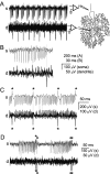
Bursts are terminated by dendritic spikes. Simultaneous extracellular recordings were made from the soma, near the axon hillock, and from the dendrites, at least 30 μm from the soma. A, Simultaneous somatic (s) and dendritic (d) recordings of spontaneous bursts in a Purkinje neuron. The diagram at the right shows the approximate location of the recording pipettes. At the end of each burst of somatic action potentials a rapid negative voltage change is seen in the dendrite. B, A single burst from the cell described in A. The dashed line shows that the peak of the dendritic spike occurs after the final somatic spike. C, Simultaneous somatic and dendritic recordings from another Purkinje neuron show that with each negative dendritic spike (denoted by asterisk), a corresponding positive potential change was recorded at the soma. Each sodium spike produced a negative potential change at the axon hillock and a corresponding positive potential change in the dendrites. The current source for the action potentials is confined to the soma, whereas the current source for the spikes that terminate the bursts is localized to the dendrites. D, Simultaneous recordings from a Purkinje neuron in which each burst of ∼25 spikes was followed immediately by a much shorter burst of 3 spikes. Both long and short bursts were terminated with a dendritic calcium spike (denoted by asterisk).
Figure 5.
Figure 5.
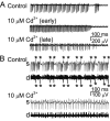
P/Q-type calcium channels are required for dendritic spikes and for bursting. A, Extracellular recordings from a Purkinje neuron under control conditions (top trace) and at 3 min (middle trace) and 10 min (bottom trace) after superfusion with cadmium (10 μm). B, Dual extracellular recordings from the soma (s) and dendrites (d) of a Purkinje neuron. Dendritic recordings show dendritic spikes as negative voltage deflections, whereas the somatic sodium spikes are positive. The polarity of the spikes is reversed in the somatic recordings. Under control conditions, each burst is terminated by a dendritic spike (denoted by asterisk). Cadmium (10 μm) inhibits dendritic but not somatic spikes (bottom traces).
Figure 6.
Figure 6.
Dendritic spikes occur spontaneously in the absence of sodium-dependent action potentials. A, Somatic (top trace) and dendritic (bottom trace) recordings before and after application of TTX (1 μm). Dendritic spikes produced positive potential changes at the soma. Each occurrence of a dendritic spike is indicated by an asterisk. B, The average firing rate (top trace) and average occurrence of dendritic calcium spikes (bottom trace) calculated every 500 msec are shown. TTX blocked firing of somatic but not dendritic spikes. Dendritic spikes continued to occur in bursts at regular intervals even in the presence of TTX. C, Average occurrence of dendritic spikes under control conditions and in TTX (left plot). The average cycle period of the bursts of calcium spikes for each cell under control conditions and in TTX is shown in the right plot. For each cell the ratio of the value measured in TTX to the value measured in control conditions was calculated for both the rate of calcium spike occurrence and cycle period. The averages of the ratios for all cells tested are shown in the histogram. Error bars represent ±SEM (n = 4, calcium spike rate; n = 8, cycle period).
Figure 7.
Figure 7.
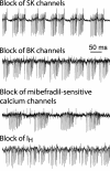
Block of SK, BK, T-type calcium, or hyperpolarization-activated channels does not prevent spontaneous bursting. Recordings of spontaneous bursting in Purkinje neurons in the presence of specific blockers for the ion channels as indicated. SK channels were blocked with apamin (100 nm), BK channels with iberiotoxin (100 nm), and hyperpolarization-activated channels with cesium (1 mm). Mibefradil (1 μm) was used to block both T- and R-type voltage-gated calcium channels. Pharmacological block of these conductances abolished neither the trimodal pattern of activity nor the generation of regular spontaneous bursts.
Figure 8.
Figure 8.
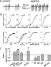
SK channels contribute to interspike and interburst intervals. A, Burst firing recorded from a Purkinje neuron under control conditions and in the presence of apamin (100 nm). B, Average cumulative probability distributions for each parameter under control conditions (open symbols) and in apamin (closed symbols) for all cells studied (mean ± SEM; n = 6). C, The ratio of the median value in apamin to the median value in control conditions was determined for each parameter in each cell tested. For each parameter, the mean of this ratio for all cells is shown. The horizontal bar indicates a ratio value of 1 corresponding to no effect of the blocker on the parameter measured. Asterisks indicate that the mean is significantly different from 1 (*p < 0.08; **p < 0.006; determined by one-way ANOVA). Error bars represent ±SEM (n = 6). The number of bursts analyzed was 2790 under control conditions and 5045 in apamin. For each cell, bursts from at least two cycles of the trimodal pattern of activity were analyzed under control conditions and in apamin.
Figure 9.
Figure 9.
Contribution of BK, T-type calcium, and hyperpolarization-activated channels to spontaneous bursting. Extracellular recordings were made from Purkinje neurons under control conditions and in the presence of specific ion channel blockers. Cumulative probability plots of burst parameters similar to those shown in Figure 8 B were made. The average of the ratio of the median value in blocker to the median value in control conditions was determined for each burst parameter. These ratios accurately reflected the changes seen in the cumulative probability plots and are shown in this figure for each blocker. The horizontal bars indicate a value of 1. Ratios that significantly differ from 1 are indicated by asterisks. A, Recordings were made under control conditions and in the presence of iberiotoxin (100 nm) to block BK channels (*p < 0.05; *p < 0.01; determined by one-way ANOVA). Error bars represent ±SEM, (n = 11). The number of bursts analyzed was 11,172 under control conditions and 8517 in iberiotoxin. B, Burst firing was recorded under control conditions and in the presence of mibefradil (1 μm) to block T-type calcium channels (*p < 0.02; *p < 0.002; determined by one-way ANOVA). Error bars represent ±SEM, (n = 5). The number of bursts analyzed was 2184 under control conditions and 3567 in mibefradil. C, The effect of mibefradil (1 μm) was examined in the continuous presence of iberiotoxin (100 nm). (*p < 0.002; **p < 0.001; determined by one-way ANOVA). Error bars represent ±SEM (n = 5). The number of bursts analyzed was 3479 in iberiotoxin alone and 2100 in iberiotoxin plus mibefradil. D, The effect of iberiotoxin (100 nm) was examined in the continuous presence of mibefradil (1 μm) (*p < 0.05; **p < 0.005; determined by one-way ANOVA). Error bars represent ±SEM (n = 6). The number of bursts analyzed was 2639 in mibefradil alone and 2329 in mibefradil plus iberiotoxin. E, Cesium (1 mm) was used to block IH. Ratios significantly different from 1 are indicated by asterisks (*p < 0.002; determined by one-way ANOVA). Error bars represent ±SEM, (n = 5). The number of bursts analyzed was 2783 under control conditions and 2864 in cesium. For each cell, all bursts from at least two cycles of the trimodal pattern of activity were analyzed under control conditions and at least two cycles in the presence of the channel blocker.

References

    1. Bal T, McCormick DA (1997) Synchronized oscillations in the inferior olive are controlled by the hyperpolarization-activated cation current I(h). J Neurophysiol 77: 3145–3156. - PubMed
    1. Cavelier P, Pouille F, Desplantez T, Beekenkamp H, Bossu JL (2002) Control of the propagation of dendritic low-threshold Ca(2+) spikes in Purkinje cells from rat cerebellar slice cultures. J Physiol (Lond) 540: 57–72. - PMC - PubMed
    1. Cingolani LA, Gymnopoulos M, Boccaccio A, Stocker M, Pedarzani P (2002) Developmental regulation of small-conductance Ca2+-activated K+ channel expression and function in rat Purkinje neurons. J Neurosci 22: 4456–4467. - PMC - PubMed
    1. Crepel F, Penit-Soria J (1986) Inward rectification and low threshold calcium conductance in rat cerebellar Purkinje cells. An in vitro study. J Physiol (Lond) 372: 1–23. - PMC - PubMed
    1. Del Negro CA, Hsiao CF, Chandler SH (1999) Outward currents influencing bursting dynamics in guinea pig trigeminal motoneurons. J Neurophysiol 81: 1478–1485. - PubMed

Publication types

MeSH terms

Substances

LinkOut - more resources