. 2004 Apr 7;24(14):3543-53.
doi: 10.1523/JNEUROSCI.5048-03.2004.

Identification and characterization of heterogeneous neuronal injury and death in regions of diffuse brain injury: evidence for multiple independent injury phenotypes

Affiliations

Identification and characterization of heterogeneous neuronal injury and death in regions of diffuse brain injury: evidence for multiple independent injury phenotypes

Richard H Singleton et al. J Neurosci. .

Abstract

Diffuse brain injury (DBI) is a consequence of traumatic brain injury evoked via rapid acceleration-deceleration of the cranium, giving rise to subtle pathological changes appreciated best at the microscopic level. DBI is believed to be comprised by diffuse axonal injury and other forms of diffuse vascular change. The potential, however, that the same forces can also directly injure neuronal somata in vivo has not been considered. Recently, while investigating DBI-mediated perisomatic axonal injury, we identified scattered, rapid neuronal somatic necrosis occurring within the same domains. Moving on the premise that these cells sustained direct somatic injury as a result of DBI, we initiated the current study, in which rats were intracerebroventricularly infused with various high-molecular weight tracers (HMWTs) to identify injury-induced neuronal somatic plasmalemmal disruption. These studies revealed that DBI caused immediate, scattered neuronal somatic plasmalemmal injury to all of the extracellular HMWTs used. Through this approach, a spectrum of neuronal change was observed, ranging from rapid necrosis of the tracer-laden neurons to little or no pathological change at the light and electron microscopic level. Parallel double and triple studies using markers of neuronal degeneration, stress, and axonal injury identified additional injured neuronal phenotypes arising in close proximity to, but independent of, neurons demonstrating plasmalemmal disruption. These findings reveal that direct neuronal somatic injury is a component of DBI, and diffuse trauma elicits a heretofore-unrecognized multifaceted neuronal pathological change within the CNS, generating heterogeneous injury and reactive alteration within both axons and neuronal somata in the same domains.


Figures

Figure 1.
Figure 1.
Passage of the HRP tracer though the brain parenchymal extracellular space. A, Coronal brain section from a rat that was not infused with HRP. At 2 hr after sham injury (4 hr after infusion), the tracer extends into the deeper layers of the neocortex and also encompasses the majority of the hippocampus (B). At 6 hr after sham injury (8 hr after infusion), the tracer has diffused throughout the majority of the brain parenchyma (C). By 24 hr after sham injury (26 hr after infusion), extracellular HRP is not readily seen, most likely because of its clearance from the CSF (D).
Figure 2.
Figure 2.
Neurons in multiple loci demonstrate altered plasmalemmal permeability to HRP and dex40 after traumatic brain injury. In animals infused with HRP and subjected to sham injury (A, neocortex), no neuronal flooding is observed, although HRP is seen in relation to the vasculature (arrowheads). In contrast, injured animals demonstrate dramatic neuronal HRP flooding in multiple sites at 2 hr after injury (C, neocortex). Flooded neuronal somata are also visible at 6 hr after injury scattered throughout these same anatomic loci, whereas at 24 hr after injury, HRP-positive neurons were few. Similarly, in sham-injured animals infused before injury with dex40, no neuronal flooding is observed in the neocortex, the CA1 region of the hippocampus (B), or any other brain region. In contrast, 2 hr after TBI, scattered neocortical neurons demonstrate striking dex40 labeling within somatic and dendritic domains. Similar findings are noted in scattered CA1 pyramidal neurons (D), as well as in other regions of the hippocampus and dentate gyrus. At a higher magnification, microvacuolization, suggestive of mitochondrial swelling, can be noted in the dendrites of dex40-flooded neurons (D, inset, arrows). Scattered neurons flooded with dex40 remained visible in the same anatomic loci at 6 and 24 hr after injury. Scale bars: A–D, 100 μm; D, inset, 25 μm.
Figure 5.
Figure 5.
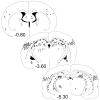
Extent and relative burden of neuronal somata demonstrating plasmalemmal injury and flooding with dex10 2 hr after moderate central fluid percussion injury. Coronal brain levels rostral (–0.80 mm relative to bregma), at the level of (–3.60 mm), and caudal (–5.30 mm) to the injury site are depicted. Each dot represents five dex10-flooded neurons within the specific brain region. As can be observed, most of the flooded neurons were noted at the level of the injury and in more caudal sites; less damage was noted in more rostral brain regions. In addition, no dex10-flooded neurons were observed directly under the injury site (–3.60 mm, dorsal midline neocortex).
Figure 3.
Figure 3.
Neurons flooded with HRP demonstrate various forms of pathological change. In injured animals surviving for 2 hr after injury, one can identify scattered HRP-flooded neurons demonstrating overt necrosis at the LM level. One such neuronal soma is shown in A, with disruption of the normal neuronal profile, pyknosis, and perinuclear vacuole formation (arrow). Also note the lucent areas in the perisomatic domain, indicative of synaptic terminal loss with glial ensheathment. The surrounding neuropil and neuroglial nuclei demonstrate little, if any, overt pathological change (arrowheads). These LM findings were confirmed at the ultrastructural level (B), with scattered HRP-flooded neurons revealing perisomatic glial ensheathment, microvacuolation, and pyknosis. In contrast to the relatively rapid cell death observed in association with permeability to HRP, other tracer-flooded neurons were found to demonstrate relatively little overt ultrastructural pathology. The neuron shown in C reveals only modest swelling of the Golgi apparatus (large arrowhead), whereas the remainder of the cytoplasm appears intact. Surrounding this neuron, one can identify HRP in the extracellular space (small arrowheads), as well as a degenerating presynaptic bouton (arrow). Scattered HRP-flooded neurons revealing intermediate levels on injury were also identified, as can be seen in D, which demonstrates an injured neuronal soma with mitochondrial swelling (arrowheads) and perisomatic glial infiltration. In all of these cases, evidence of somatic HRP influx was confirmed in adjacent unstained sections. Scale bars: A, 50 μm; B, C, 4 μm; D, 2 μm.
Figure 4.
Figure 4.
Neuronal somata demonstrate altered membrane permeability and flooding with dex10 within 5 min of injury. In this low-power image of the rat brain 5 min after moderate midline central fluid percussion injury, neurons flooded with dex10 tracer are prevalent throughout the mediodorsal neocortex (demarcated by the dotted line). The CA1 region of the hippocampus is seen at the bottom left. Inset, At a higher magnification, neuronal profiles flooded with the dex10 tracer are evident scattered among other neurons demonstrating no evidence of tracer flooding. The deep layers of the neocortex can be seen at the bottom left, whereas the more superficial cortical layers can be observed at the top right. Note the areas of very intense dex10 labeling seen in relation to the vasculature (arrowheads). Scale bars: 300 μm; inset, 200 μm.
Figure 6.
Figure 6.
Neurons flooding with HRP and dex10 reveal partial overlap. Animals were infused with HRP and dex10, subjected to moderate central FPI, allowed to survive for 2 hr, and then reacted with antibodies to HRP. In sections from multiple brain regions, including the neocortex (pictured) and the hippocampus, some neurons flooded with dex10 also demonstrated cytoplasmic HRP (arrowheads). In addition to these double-labeled cells, however, a second neuronal population was observed to flood only with HRP but remained impermeable to the dextran tracer (arrows). Those neurons observed to flood with both HRP and dex10 appear overtly pathological, with irregular profiles, whereas other neurons flooding with HRP alone do not demonstrate any other pathology at the LM level. Scale bars, 100 μm.
Figure 7.
Figure 7.
Neuronal HSP70 expression is induced after diffuse traumatic brain injury. No HSP70 expression is evident in sham-injured animals. At 2 hr after injury, no upregulation of HSP70 is noted, whereas at 6 hr, scattered HSP70-positive neurons are localized in the mediodorsal neocortex and several regions of the hippocampus and dentate gyrus. Expression of this cell stress marker increased further by 24 hr after injury, at which time marked immunolabeling of scattered neuronal somata and their dendritic processes can be noted in the same regions in which HSP70 expression was seen at 6 hr, including the neocortex (pictured). Scale bars, 100 μm.
Figure 8.
Figure 8.
Neuronal degeneration or death occurs after diffuse brain injury. In rats subjected to sham injury, FJ staining reveals only low levels of background fluorescence throughout the brain (A). In contrast, brain sections from rats subjected to moderate central fluid percussion injury reveal scattered, brightly stained, FJ-positive neuronal profiles at 2, 6 (B), and 24 hr after injury in the neocortex and hippocampus and dentate gyrus. Many of these labeled neurons are overtly pathological, demonstrating irregular somatic profiles. Scale bars, 100 μm.
Figure 9.
Figure 9.
Neurons demonstrating perisomatic axotomy reveal no evidence of somatic plasmalemmal disruption. In animals infused with dex40 (green), subjected to TBI, and killed 2 hr after injury, tracer-flooded neurons (arrowheads) do not colocalize with other neurons linked to adjacent APP-positive reactive axonal swellings (red, arrow). The heterogeneous nature of diffuse injury is also depicted, in that one can observe two directly adjacent neurons manifesting distinctly different injury phenotypes, with an overtly pathological, microvacuolated, dex40-flooded neuron adjacent to an axotomized neuron demonstrating no other evidence of perturbation. Scale bar, 50 μm.
Figure 10.
Figure 10.
Reactive neuronal somatic expression of HSP70 is independent of both cell membrane permeability alteration and perisomatic axotomy. In this triple-labeled section from an animal infused with dex40 and surviving for 24 hr after injury, fluorescent labeling for dex40 (green), APP (red), and HSP70 (blue) can be observed. As noted above, no overlap occurs between those axotomized neurons linked to disconnected, APP-positive reactive axonal swellings (triple arrowhead) and neurons flooding with dex40 (arrowheads). HSP70-positive neuronal somata (double arrowhead) do not reveal any colocalization with either axotomized neurons or those neurons demonstrating altered plasmalemmal permeability. Intense non-neuronal dex40 staining can be seen, attributable to diffusion of the tracer along the perivascular sleeve (arrows). Scale bar, 50 μm.
Figure 11.
Figure 11.
Neuronal somata demonstrating permeability to HRP reveal incomplete overlap with FJ. This section from an animal surviving for 2 hr after injury demonstrates a lack of complete concordance between these two markers, with some HRP-flooded neurons (red) in the neocortex staining positively for FJ (green; arrow) and others revealing no evidence of FJ labeling (arrowheads). Scale bars, 100 μm.

Similar articles

Cited by

References

    1. Abravaya K, Myers MP, Murphy SP, Morimoto RI (1992) The human HSP hsp70 interacts with HSF, the transcription factor that regulates heat shock gene expression. Genes Dev 6: 1153–1164. - PubMed
    1. Adams JH (1992) Head injury. In: Greenfield's neuropathology (Adams JH, Duchen LW, eds), pp 106–152. New York: Oxford UP.
    1. Adams JH, Mitchell DE, Graham DI, Doyle D (1977) Diffuse brain damage of immediate impact type: its relationship to “primary brain-stem damage” in head injury. Brain 100: 489–502. - PubMed
    1. Barros LF, Castro J, Bittner CX (2002) Ion movements in cell death: from protection to execution. Biol Res 35: 209–214. - PubMed
    1. Blumbergs PC, Scott G, Manavis J, Wainwright H, Simpson DA, McLean AJ (1994) Staining of amyloid precursor protein to study axonal damage in mild head injury. Lancet 344: 1055–1056. - PubMed

Publication types

MeSH terms

LinkOut - more resources