. 2004 Apr 7;24(14):3592-9.
doi: 10.1523/JNEUROSCI.5167-03.2004.

Oligomerization of Alzheimer's beta-amyloid within processes and synapses of cultured neurons and brain

Affiliations

Oligomerization of Alzheimer's beta-amyloid within processes and synapses of cultured neurons and brain

Reisuke H Takahashi et al. J Neurosci. .

Abstract

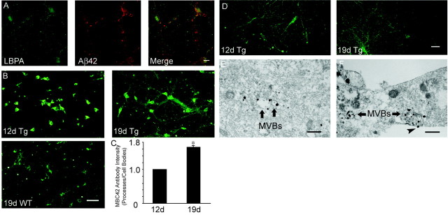
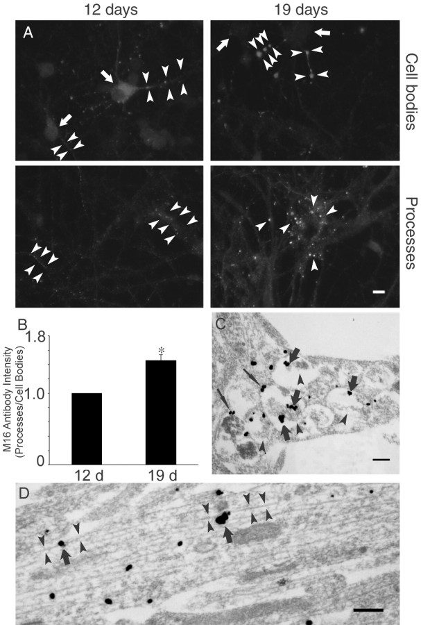
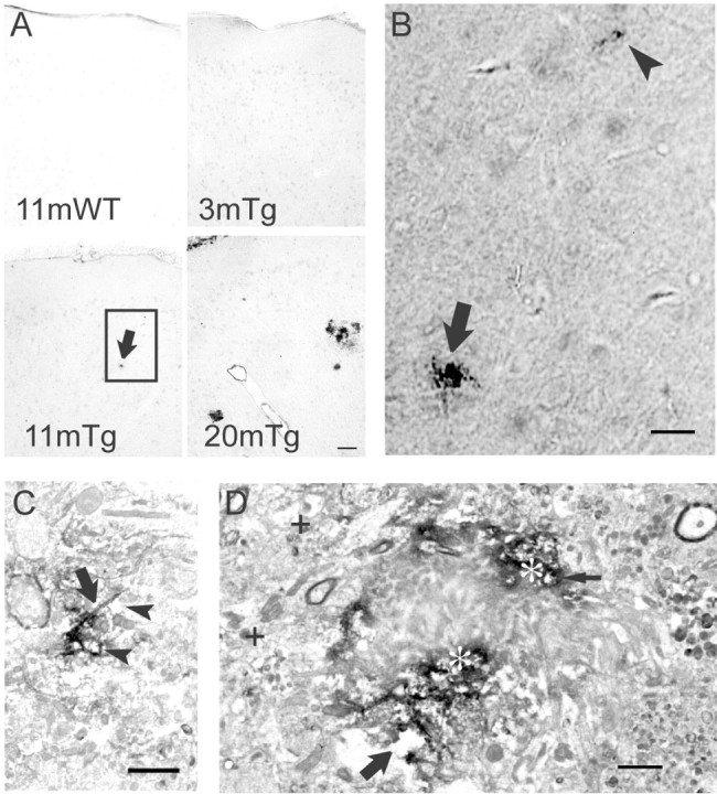
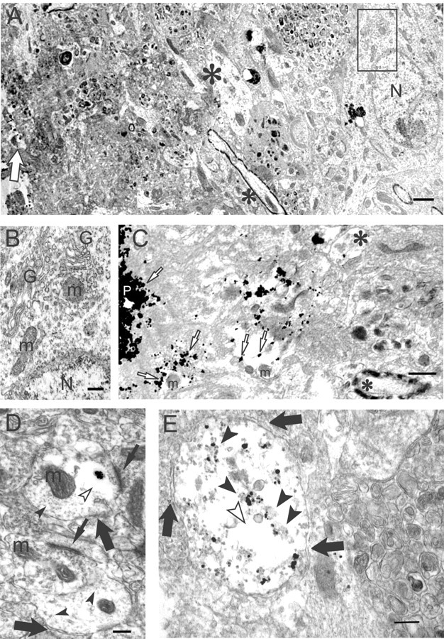
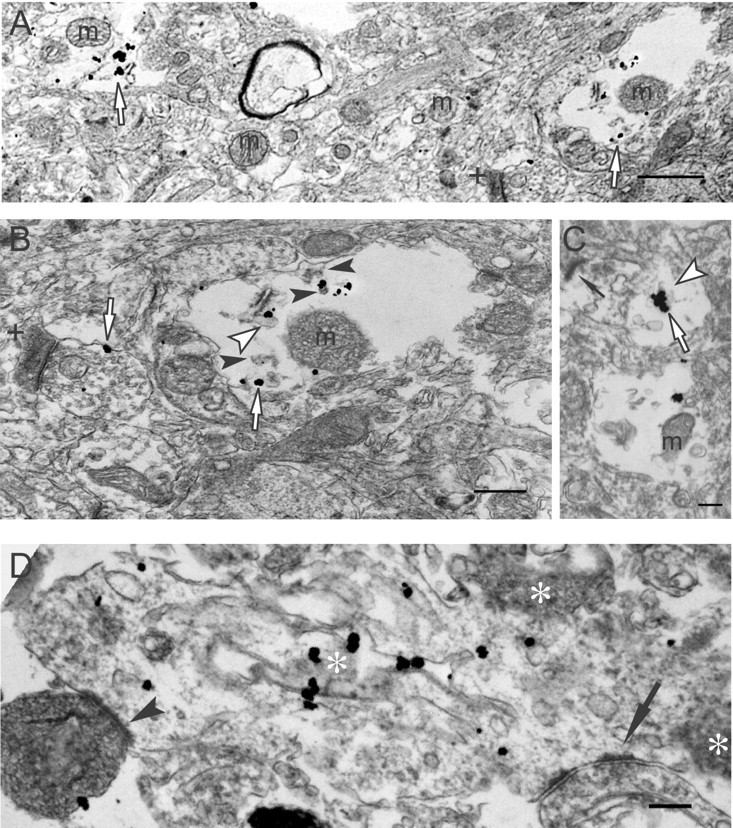
Multiple lines of evidence implicate beta-amyloid (Abeta) in the pathogenesis of Alzheimer's disease (AD), but the mechanisms whereby Abeta is involved remain unclear. Addition of Abeta to the extracellular space can be neurotoxic. Intraneuronal Abeta42 accumulation is also associated with neurodegeneration. We reported previously that in Tg2576 amyloid precursor protein mutant transgenic mice, brain Abeta42 localized by immunoelectron microscopy to, and accumulated with aging in, the outer membranes of multivesicular bodies, especially in neuronal processes and synaptic compartments. We now demonstrate that primary neurons from Tg2576 mice recapitulate the in vivo localization and accumulation of Abeta42 with time in culture. Furthermore, we demonstrate that Abeta42 aggregates into oligomers within endosomal vesicles and along microtubules of neuronal processes, both in Tg2576 neurons with time in culture and in Tg2576 and human AD brain. These Abeta42 oligomer accumulations are associated with pathological alterations within processes and synaptic compartments in Tg2576 mouse and human AD brains.


Figures

Figure 1.
Figure 1.
Aβ42 within cultured neurons localizes to late endosomal vesicles and increases with time in culture, especially within processes. A, Confocal immunofluorescence microscopy of primary neurons at 19 d in culture. Aβ42 fluorescence, with the Chemicon Aβ42 Ab, partially localized with LBPA, which is known to be associated especially with late endosomes. Scale bar, 10 μm. B, Immunofluorescence of Tg2576 mouse primary neurons stained with mAb MBC42 against the C terminus of Aβ42. Note the Aβ42 increases in Tg2576 (Tg) neurons between 12 d (left) and 19 d (right) in culture. There is less Aβ42 immunofluorescence in wild-type (WT) neurons cultured for 19 d (below). Scale bar, 50 μm. C, Tg2576 primary neurons: ratio of Aβ42 immunofluorescence intensity in neuronal processes divided by that in cell bodies (processes/cell bodies) between 12 and 19 d in culture, with the ratio standardized to 1.0 for 12 d in culture. The Aβ42 ratio of processes/cell bodies increased 1.65 ± 0.06-fold between 12 and 19 d in culture; p < 0.001. D, Representative images of α-tubulin immunofluorescence in Tg2576 mouse primary neurons at 12 and 19 d in culture. There was no significant increase inα-tubulin fluorescence, compared with that of Aβ42 fluorescence (B), within processes between 12 and 19 d in culture. Scale bar, 50 μm. E, Cell body Aβ42 detected by immunogold electron microscopy using Ab MBC42. Representative Aβ42 in the outer membrane of a MVB, containing characteristic internal vesicles, in the cell body of a Tg2576 primary neuron at 21 d in culture. Scale bar, 500 nm. F, Aβ42 immuno-EM of a neurite process. Representative image of Aβ42 accumulation in outer membranes and rarely inner vesicles of MVBs in a neurite process of a Tg2576 primary neuron at 21 d in culture, detected by immunogold EM with MBC42. Note also an Aβ42 gold particle at the plasma membrane (arrowhead). Scale bar, 500 nm.
Figure 2.
Figure 2.
Biochemical analysis of intraneuronal Aβ accumulation in primary neurons with time in culture. A, Increased levels of Aβ with time in culture in primary neurons from Tg2576 mouse by immunoprecipitation–Western blot. Equal amounts of 12- and 19-d-old cultured neuron cell lysates were immunoprecipitated with 4G8 and immunoblotted with 6E10 for determination of Aβ and full-length APP levels. After immunoprecipitation with 4G8, supernatants were immunoblotted with antibodies against NeuN or α-tubulin. B, Aβ levels increased 3.36 ± 0.51-fold from 12 to 19 d in culture (p < 0.03). A slight increase in APP levels (1.46 ± 0.29-fold) from 12 to 19 d in culture was not statistically significant (p = 0.19). Data are expressed as mean ± SEM of three independent experiments. C, Aggregation-state specificity of antibodies against Aβ42. Aggregated Aβ42 (200 ng) was separated by gel electrophoresis, transferred to polyvinylidene-difluoride membrane, and blotted with MBC42, 6E10, and M16. MBC42 detected especially Aβ42 monomers and some dimers but did not recognize oligomers. 6E10 recognized most aggregation states of Aβ42, being most sensitive for monomers. M16 mainly recognized larger Aβ42 oligomers, with relatively minor reactivity to Aβ monomers.
Figure 3.
Figure 3.
Aβ42 oligomers accumulate within neuronal processes with time in culture. A, Increased oligomeric Aβ42 (M16) immunofluorescence in processes of Tg2576 primary neurons with time in culture. Top panel, Localization of Aβ42 in cell bodies (arrows) and processes (arrowheads) at 12 d (left) and 19 d (right) in culture. Bottom panel, Localization of Aβ42 oligomers in neuronal processes. Scale bar, 10 μm. B, Ratio of oligomeric Aβ42 immunofluorescence intensity in processes to that in cell bodies (processes/cell bodies) at 12 and 19 d in culture in Tg2576 primary neurons, with intensity ratio standardized as 1.0 at day 12 in culture. There was a 1.46 ± 0.09-fold increase in the process-to-cell body ratio of Aβ42 oligomer fluorescence between 12 and 19 d in culture; p < 0.01. C, Aβ42 oligomer accumulation in neuronal processes of Tg2576 neurons at 19 d in culture, especially within endosomal-like vesicles (thick arrows), and less prominently in their outer membranes (thin arrows). These processes are associated with morphological alterations, including fibrillar dark material (arrowheads) that appears associated with intravesicular Aβ42 oligomers. Scale bar, 250 nm. D, Aβ42 oligomers (arrows) also localized and accumulated along microtubules (arrowheads) within neuronal processes of Tg2576 neurons at 19 d in culture. Scale bar, 250 nm.
Figure 4.
Figure 4.
Aβ42 oligomers accumulate in Tg2576 mouse brain. A, Low-magnification light microscopy images of M16 Aβ42 oligomer immunoperoxidase labeling of wild-type (WT) and Tg2576 (Tg) mouse brains at different ages. Aβ42 oligomers were not detected in 11-month-old wild-type (top left) or 3-month-old Tg2576 mouse brains (top right) but were faintly detected as dense micro-deposits (bottom left, arrow) in an 11-month-old Tg2576 mouse. By 20 months, M16 Aβ42 oligomer staining showed increased number and size of microdeposits (bottom right) and also surrounded neuritic plaques. Scale bar, 50 μm. B, High-magnification light microscopic image of the rectangle in A (bottom left), showing a microdeposit (arrow) and even smaller Aβ42 oligomer aggregation before plaques in an 11-month-old Tg2576 mouse (arrowhead). Scale bar, 30 μm. C, Immunoperoxidase EM of Aβ42 oligomer aggregations distant from an obvious plaque. Aβ42 oligomer immunoreactivity appears concentrated along processes (arrow), and structures around the aggregation sites are degenerating (vacuolated spaces, arrowheads). Scale bar, 1μm. D, Ultrastructural localization of Aβ42 oligomers around a plaque. Marked accumulation of immunoperoxidase reaction product (appearing black; asterisks) representing Aβ42 oligomers surrounds an unlabeled plaque core. Areas around the plaque core are degenerated and include degenerating neurites (thin arrow) and abnormal empty spaces (large arrow). For reference, more normal neurites distant from the plaque are also seen (plus signs). Scale bar, 1 μm.
Figure 5.
Figure 5.
Aβ oligomers by immunogold EM accumulate in dystrophic processes and synapses of Tg2576 mouse brain. A, A lower-magnification image of a degenerated, morphologically abnormal area around a plaque core (left) surrounded by normal-appearing neuropil (right). A degenerating neurite (empty arrow) is observed close to the plaque at the left edge of the figure. Normal dendrite (large asterisk) and a myelinated axon (small asterisk) are at the border of the normal and abnormal areas. A normal neuron is seen distant from the plaque (right); the rectangle within this normal-appearing neuron is enlarged in B, below. N, Nucleus. Scale bar, 1 μm. B, Normal organelles and cytoarchitecture can be seen in this higher-magnification image of the small rectangle within the neuron in A. G, Golgi apparatus; m, mitochondrion; N, nucleus. Scale bar, 250 nm. C, A higher-magnification image of an Aβ oligomer containing degenerating neurites and abnormal empty spaces just around a plaque core (P). Aβ oligomers (empty arrow) are associated with the plaque core, and degenerating neurites are associated with empty spaces. A relatively normal dendrite with an isolated M16 gold particle (large asterisk) and a myelinated axon (small asterisk) are observed distant from the plaque core. m, Mitochondrion. Scale bar, 500 nm. D, A cluster of gold particles representing Aβ42 oligomers in a postsynaptic profile near an active zone of a synapse (top thin arrow). The plasma membranes of two postsynaptic profiles are indicated (large arrows). The cluster of Aβ42 oligomers is surrounded by a clear area (empty arrowhead), in which the normal microtubular network (filled arrowheads) is absent. The lower synaptic profile, lacking Aβ42 oligomers, shows a normal microtubular network throughout. Synaptic active zones are indicated by thin arrows. m, Mitochondrion. Scale bar, 250 nm. E, M16 immunogold EM of a Tg2576 neuritic process. Vertical section through a neuritic process in an 18-month-old Tg2576 mouse brain. Distant from a plaque, substantial Aβ42 oligomer gold particles are associated with granular and fibril-like electron-dense material (arrowheads) in an abnormal pathological-appearing process with poorly defined cytoarchitecture, including empty spaces (empty arrowhead) and lacking any normal organelles, such as mitochondria. Arrows outline the plasma membrane of the neuritic process. Scale bar, 250 nm.
Figure 6.
Figure 6.
Aβ42 oligomerization within dystrophic processes of human AD brain. A, Aβ42 oligomers (empty arrows) are observed in degenerating neurites and are not associated with more normal-appearing neuropil (middle area of image). A darkened, abnormal-appearing postsynaptic profile is also observed (plus sign). m, Mitochondrion. Scale bar, 1μm. B, A higher-power image of a degenerating neurite in A (A, right empty arrow). Aβ42 oligomers (empty arrows) are associated with a tubulovesicular-like organelle (empty arrowhead) and granular fibril-like electron-dense material (filled arrowheads). Isolated Aβ42 oligomers localize to the membrane of presynaptic profile (left empty arrow) associated with a darkened, degenerating-appearing postsynaptic profile (plus sign). m, Mitochondrion. C, Aggregated Aβ42 oligomers (empty arrow) appear associated with a tubulovesicular-like organelle (empty arrowhead) in a postsynaptic compartment with an active zone (thin arrow). m, Mitochondrion. Scale bar, 300 nm. D, M16 Aβ42 oligomers aggregate on tubular-like membranous structures within a disrupted, swollen dendrite containing abnormal dark profiles (asterisks), consistent with degeneration; no normal organelles can be found in this postsynaptic profile. Presynaptic profiles are seen on the right (arrow) and left (arrowhead) corners of the image; the left one is darkened, indicative of degeneration. Scale bar, 250 nm.

References

    1. Busciglio J, Pelsman A, Wong C, Pigino G, Yuan ML, Mori H, Yankner BA (2002) Altered metabolism of the amyloid beta precursor protein is associated with mitochondrial dysfunction in Down's syndrome. Neuron 33: 677–688. - PubMed
    1. Cataldo AM, Peterhoff CM, Troncoso JC, Gomez-Isla T, Hyman BT, Nixon RA (2000) Endocytic pathway abnormalities precede amyloid beta deposition in sporadic Alzheimer's disease and Down syndrome: differential effects of APOE genotype and presenilin mutations. Am J Pathol 157: 277–286. - PMC - PubMed
    1. Chan J, Aoki C, Pickel VM (1990) Optimization of differential immunogold-silver and peroxidase labeling with maintenance of ultrastructure in brain sections before plastic embedding. J Neurosci Methods 33: 113–127. - PMC - PubMed
    1. Chapman PF, White GL, Jones MW, Cooper-Blacketer D, Marshall VJ, Irizarry M, Younkin L, Good MA, Bliss TV, Hyman BT, Younkin SG, Hsiao KK (1999) Impaired synaptic plasticity and learning in aged amyloid precursor protein transgenic mice. Nat Neurosci 2: 271–276. - PubMed
    1. D'Andrea MR, Nagele RG, Wang HY, Peterson PA, Lee DH (2001) Evidence that neurones accumulating amyloid can undergo lysis to form amyloid plaques in Alzheimer's disease. Histopathology 38: 120–134. - PubMed

Publication types

MeSH terms