. 2004 Apr 20;101(16):5886-91.
doi: 10.1073/pnas.0308456101. Epub 2004 Apr 7.

Genetic evidence for nonredundant functional cooperativity between NPC1 and NPC2 in lipid transport

Affiliations

Genetic evidence for nonredundant functional cooperativity between NPC1 and NPC2 in lipid transport

David E Sleat et al. Proc Natl Acad Sci U S A. .

Abstract

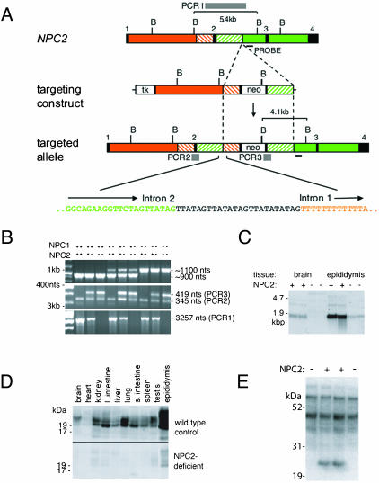
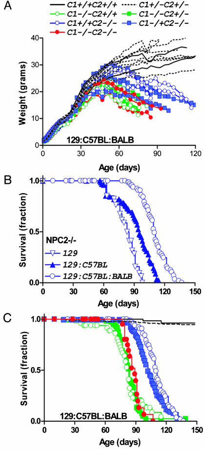
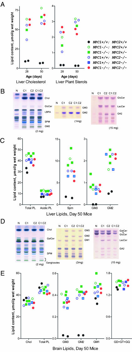
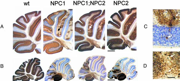
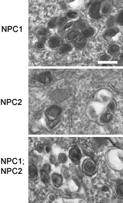
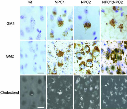
Niemann-Pick C (NPC) disease is a fatal neurodegenerative disorder characterized by a lysosomal accumulation of cholesterol and other lipids within the cells of patients. Clinically identical forms of NPC disease are caused by defects in either of two different proteins: NPC1, a lysosomal-endosomal transmembrane protein and NPC2, a soluble lysosomal protein with cholesterol binding properties. Although it is clear that NPC1 and NPC2 are required for the egress of lipids from the lysosome, the precise roles of these proteins in this process is unknown. To gain insight into the normal function of NPC2 and to investigate its interactions, if any, with NPC1, we have generated a murine NPC2 hypomorph that expresses 0-4% residual protein in different tissues and have examined its phenotype in the presence and absence of NPC1. The phenotypes of NPC1 and NPC2 single mutants and an NPC1;NPC2 double mutant are similar or identical in terms of disease onset and progression, pathology, neuronal storage, and biochemistry of lipid accumulation. These findings provide genetic evidence that the NPC1 and NPC2 proteins function in concert to facilitate the intracellular transport of lipids from the lysosome to other cellular sites.


Figures

Fig. 1.
Fig. 1.
Targeted disruption of the murine NPC2 gene (NPC2). (A) Structure of NPC2, the targeting construct, the targeted locus and the sequence at the NPC2 intron 2:left arm intron 1 junction. BglII sites; boxes: black shading, exons; unshaded, thymidine kinase (tk) and neo; orange, intron 1; green, introns 2 and 3; green and orange dashed, sequences duplicated in the targeted allele. The strategy for inactivating NPC2 was to insert a loxP-flanked neo cassette into exon 2 with the concomitant replacement of amino acid cysteine 42 with a stop codon and a deletion of amino acids 43–53. An aberrant recombination event, characterized by Southern blotting and sequencing of genomic PCR products, resulted in the insertion of the neo selection marker and part of the left arm containing intron 1 into intron 2 of NPC2. Twenty-four nucleotides of unknown derivation are inserted at the site of the intron 2 – intron 1 junction. Gray bars depict informative PCR products PCR1, PCR2, and PCR3 for NPC2 genotyping. (B) PCR genotyping of genomic mouse tail DNA samples representing all combinations of NPC1 and NPC2 genotype. (C) Northern blot analysis of total RNA purified from the brain and epididymis of 30-day-old wild-type and homozygous NPC2-targeted mice. (D) Immunodetection of NPC2 in different tissues from 30-day wild-type and homozygous NPC2-targeted mice. (E) Mannose-6-phosphate containing glycoproteins in epididymal extracts detected with 125I-labeled cation-independent mannose 6-phosphate receptor in a blot assay.
Fig. 2.
Fig. 2.
Growth and survival of NPC2 hypomorph mice in the presence or absence of NPC1. (A) Growth of male NPC mutant mice. (B) Survival of NPC2 hypomorph mice in different genetic backgrounds. (C) Survival of NPC2 hypomorph mice in the presence or absence of NPC1. Symbols are as in A; control, n = 11 (two NPC1+/+;NPC2+/+, nine NPC1+/–;NPC2+/–); NPC1 mutant n = 7 (five NPC1–/–;NPC2+/– and two NPC1+/+); NPC2 mutant n = 6 (three NPC1+/+;NPC2–/– and three NPC1+/–;NPC2–/–) and NPC1-NPC2 double mutant, n = 3.
Fig. 3.
Fig. 3.
Lipid accumulation in NPC mutant mice. (A) Liver cholesterol and dietary plant sterols (campesterol, sitosterol, and sitostanol) determined by gas chromatography at 28 and 50 days. (B and C) Thin-layer chromatography (TLC) and quantitation of lipids in liver. (D and E) TLC and quantitation of lipids in brain. (B and D) Left to right: main lipids, gangliosides, neutral glycolipids. Note: in the TLC of neutral glycolipids, GalCer resolves into two bands depending on fatty acid composition and in D; the upper GalCer band and GlcCer are not resolved. The tissue weight equivalent is indicated below. Wt, wild type control; C1, C2, and C1;C2, NPC1, NPC2, and NPC1;NPC2 mutant mice, respectively. Chol, cholesterol; GlcCer, glucosylceramide; LBPA, lyso(bis)phosphatidic acid; SPM, sphingomyelin; LacCer, lactosylceramide; GalCer, galactosylceramide; GD+GT+GQ, di-, tri-, and tetrasialogangliosides. Mice were in a mixed C57BL6/129SvEv/BALBc background.
Fig. 4.
Fig. 4.
Neurodegenerative changes affecting cerebellar Purkinje cells. Calbindin-stained sections of cerebellum from wild-type (Wt), NPC1, NPC1;NPC2, and NPC2 mutant mice at (A) 7 and (B) 11 wk of age. (C) A calbindin-stained Purkinje cell in an NPC2 hypomorph mouse cerebellum showing neurodegenerative changes (dendritic and axonal swellings, upper and lower arrows, respectively) typical of all three disease models. (D) Parvalbumin–immunoreactive axonal enlargements (spheroids) in the deep white matter of a cerebellar folium (arrows) of an NPC2 hypomorph mouse that are typical for all three models. Nissl counterstain. Mice were in a mixed C57BL6/129SvEv/BALBc background. [Bars = 500 μm(A), 1,100 μm(B), and 80 μm(D).]
Fig. 5.
Fig. 5.
Ultrastructure of neuronal storage material by electron microscopy. (Bar = 0.25 μm.) Mice were 7 wk old and in a mixed C57BL6/129SvEv/BALBc background.
Fig. 6.
Fig. 6.
Lipid storage in NPC mutants. Examples of individual neurons shown by * over nucleus. Mice were 7 wk old and in a mixed C57BL6/129SvEv/BALBc background. [Bars = 25 μm (GM2 and GM3 ganglioside immunocytochemistry) and 15 μm (cholesterol detection by filipin staining)].

References

    1. Ioannou, Y. A. (2001) Nat. Rev. Mol. Cell Biol. 2, 657–668. - PubMed
    1. Davies, J. P., Chen, F. W. & Ioannou, Y. A. (2000) Science 290, 2295–2298. - PubMed
    1. Okamura, N., Kiuchi, S., Tamba, M., Kashima, T., Hiramoto, S., Baba, T., Dacheux, F., Dacheux, J. L., Sugita, Y. & Jin, Y. Z. (1999) Biochim. Biophys. Acta 1438, 377–387. - PubMed
    1. Zervas, M., Somers, K. L., Thrall, M. A. & Walkley, S. U. (2001) Curr. Biol. 11, 1283–1287. 86. - PubMed
    1. Friedland, N., Liou, H.-L., Lobel, P. & Stock, A. M. (2003) Proc. Natl. Acad. Sci. USA 100, 2512–2517. - PMC - PubMed

Publication types

MeSH terms

LinkOut - more resources