Review
. 2004 Apr 21;23(8):1681-7.
doi: 10.1038/sj.emboj.7600186. Epub 2004 Apr 8.

Cullin-based ubiquitin ligases: Cul3-BTB complexes join the family

Affiliations
Review

Cullin-based ubiquitin ligases: Cul3-BTB complexes join the family

Lionel Pintard et al. EMBO J. .

Erratum in

  • EMBO J. 2005 Mar 9;24(5):1092

Abstract

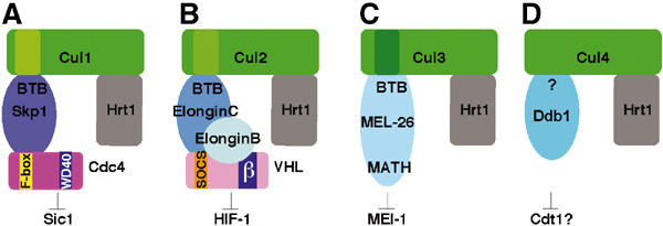
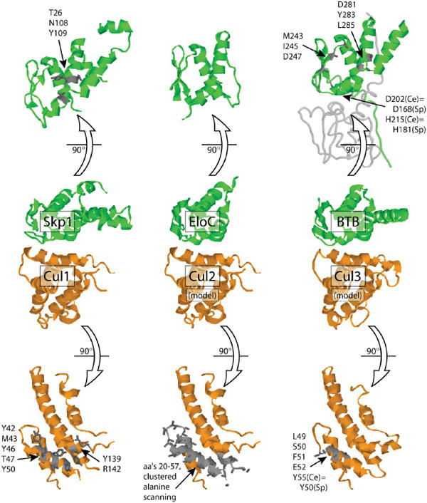
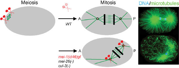
Cullin-based E3 ligases target substrates for ubiquitin-dependent degradation by the 26S proteasome. The SCF (Skp1-Cul1-F-box) and ECS (ElonginC-Cul2-SOCS box) complexes are so far the best-characterized cullin-based ligases. Their atomic structure has been solved recently, and several substrates have been described in different organisms. In addition to Cul1 and Cul2, higher eucaryotic genomes encode for three other cullins: Cul3, Cul4, and Cul5. Recent results have shed light on the molecular composition and function of Cul3-based E3 ligases. In these complexes, BTB-domain-containing proteins may bridge the cullin to the substrate in a single polypeptide, while Skp1/F-box or ElonginC/SOCS heterodimers fulfill this function in the SCF and ECS complexes. BTB-containing proteins are evolutionary conserved and involved in diverse biological processes, but their function has not previously been linked to ubiquitin-dependent degradation. In this review, we present these new findings and compare the composition of Cul3-based ligases to the well-defined SCF and ECS ligases.


Figures

Figure 1
Figure 1
Cullin-based ligases display striking similarities. The known cullin-based ligases including the recently identified Cul3-based E3 ligases display similar overall composition and structure. In SCF (A) and ECS complexes (B), Skp1 and ElonginC bridge the interaction between the cullin and the substrate-recognition protein (F-box and SOCS-box proteins, respectively). These adaptors bind substrates through a distinct protein–protein interaction domain (e.g. WD40 in the case of the F-box protein Cdc4, and β-domain in the case of VHL). In Cul3-based complexes (C), BTB proteins incorporate features of Skp1/F-box or ElonginC/SOCS-box dimers, and are thought to bridge cullin and the substrate in a single polypeptide (e.g. MEL-26 bridges the interaction between CUL-3 and MEI-1 in C. elegans). (D) Cul4 has recently been implicated in the control of DNA replication and DNA repair in C. elegans, humans, and S. pombe. While no interaction of Cul4 with Skp1, ElonginC, or BTB proteins could be observed, it was shown recently that Cul4 binds Ddb1. It is thus possible that Cul4-like ligases may use a novel adaptor family to interact with its substrates (Wertz et al, 2004).
Figure 2
Figure 2
Structural analysis of the cullin–BTB interaction. The CUL-1/SKP1 co-crystal structure (Zheng et al, 2002) served as the basis to model the interface between CUL-2 and CUL-3 with human ElonginC (Stebbins et al, 1999) and C. elegans MEL-26 (based on the human PLZF BTB domain structure (Ahmad et al, 1998). The highlighted residues have been shown by mutagenesis to be important for the cullin–BTB interaction (Pause et al, 1999; Zheng et al, 2002; Pintard et al, 2003; Geyer et al, 2003; Xu et al, 2003). ‘Sp' indicates corresponding positions in S. pombe Btb3 and Pcu3. Note that two mutations in Btb3 that disrupt its interaction with Pcu3 (D168A, H181L) are not found at the predicted BTB/cullin interface but instead are located near the putative BTB/BTB dimer interface as seen in PLZF.
Figure 3
Figure 3
The CUL-3-based complex is required for MEI-1 degradation at the meiosis-to-mitosis transition in C. elegans embryos. In C. elegans embryos, two different microtubule-based structures assemble within a 20 min interval in the same cytoplasm, for example, the meiotic and the mitotic spindle. Contrary to the anastral meiotic spindle that forms close to the cell cortex, the mitotic spindle assembles and elongates along the antero-posterior axis of the embryo (A–P). Long arrays of astral microtubules and the formation of a robust spindle are required during mitosis for correct spindle positioning, elongation, and cytokinesis. The microtubule-severing complex MEI-1/2 is essential for formation of the meiotic spindle (Clark-Maguire and Mains, 1994a, 1994b; Srayko et al, 2000). However, inactivation of this complex through MEI-1 (in red) degradation by the CUL-3-based ligase is a prerequisite for the assembly of a functional mitotic spindle. The regulation of MEI-1 degradation is not known.

References

    1. Ahmad KF, Engel CK, Prive GG (1998) Crystal structure of the BTB domain from PLZF. Proc Natl Acad Sci USA 95: 12123–12128 - PMC - PubMed
    1. Bomont P, Cavalier L, Blondeau F, Ben Hamida C, Belal S, Tazir M, Demir E, Topaloglu H, Korinthenberg R, Tuysuz B, Landrieu P, Hentati F, Koenig M (2000) The gene encoding gigaxonin, a new member of the cytoskeletal BTB/kelch repeat family, is mutated in giant axonal neuropathy. Nat Genet 26: 370–374 - PubMed
    1. Clark-Maguire S, Mains PE (1994a) Localization of the mei-1 gene product of Caenorhabditis elegans, a meiotic-specific spindle component. J Cell Biol 126: 199–209 - PMC - PubMed
    1. Clark-Maguire S, Mains PE (1994b) mei-1, a gene required for meiotic spindle formation in Caenorhabditis elegans, is a member of a family of ATPases. Genetics 136: 533–546 - PMC - PubMed
    1. Deffenbaugh AE, Scaglione KM, Zhang L, Moore JM, Buranda T, Sklar LA, Skowyra D (2003) Release of ubiquitin-charged Cdc34-S-Ub from the RING domain is essential for ubiquitination of the SCF(Cdc4)-bound substrate Sic1. Cell 114: 611–622 - PubMed

Publication types

MeSH terms