. 2004 Apr 21;23(8):1845-56.
doi: 10.1038/sj.emboj.7600195. Epub 2004 Apr 8.

Hgc1, a novel hypha-specific G1 cyclin-related protein regulates Candida albicans hyphal morphogenesis

Affiliations

Hgc1, a novel hypha-specific G1 cyclin-related protein regulates Candida albicans hyphal morphogenesis

Xinde Zheng et al. EMBO J. .

Abstract

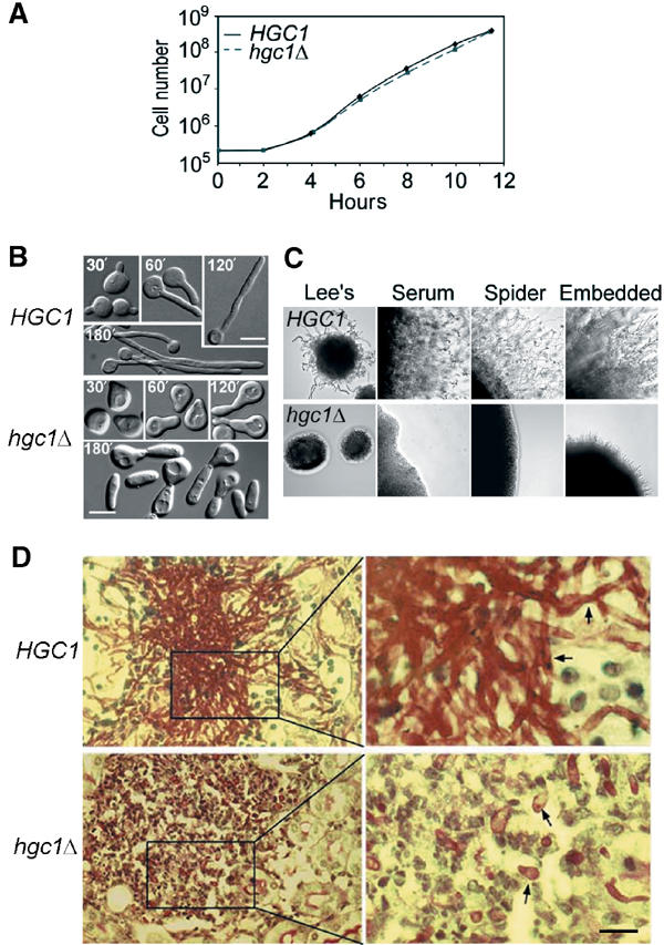
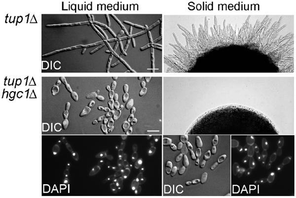
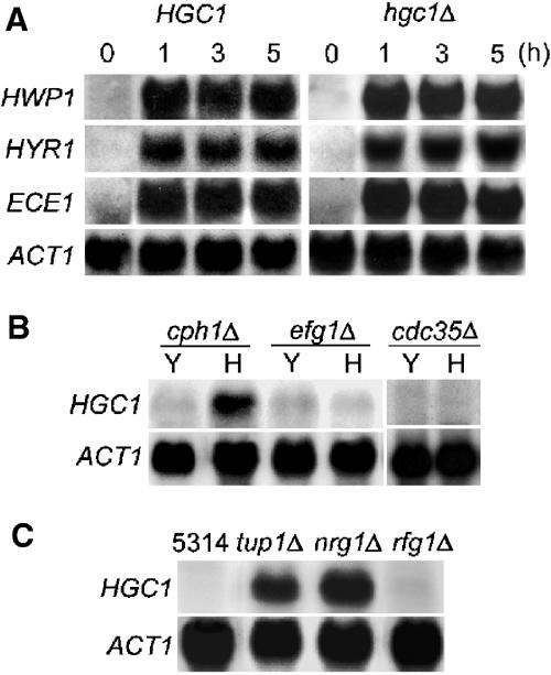
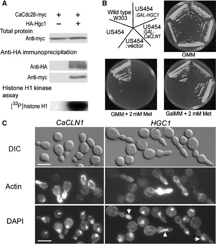
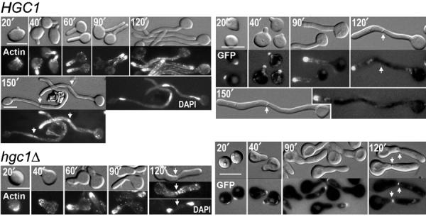
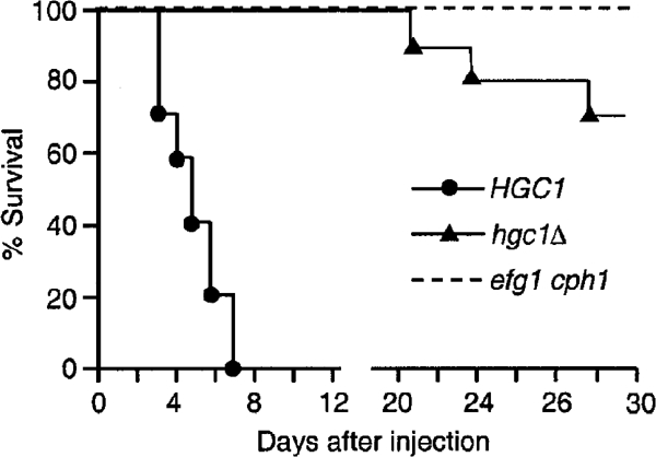
The human fungal pathogen Candida albicans switches from yeast to hyphal growth when exposed to serum or phagocytosed. However, the importance of this morphological switch for virulence remains highly controversial due to the lack of a mutant that affects hyphal morphogenesis only. Although many genes specifically expressed in hyphal cells have been identified and shown to encode virulence factors, none is required for hyphal morphogenesis. Here we report the first hypha-specific gene identified, HGC1, which is essential for hyphal morphogenesis. Deletion of HGC1 abolished hyphal growth in all laboratory conditions tested and in the kidneys of systemically infected mice with markedly reduced virulence. HGC1 expression is co-regulated with other virulence genes such as HWP1 by the cAMP/protein kinase A signaling pathway and transcriptional repressor Tup1/Nrg1. Hgc1 is a G1 cyclin-related protein and co-precipitated with the cyclin-dependent kinase (Cdk) CaCdc28. It has recently emerged that cyclin/Cdk complexes promote other forms of polarized cell growth such as tumor cell migration and neurite outgrowth. C. albicans seems to have adapted a conserved strategy to control specifically hyphal morphogenesis.


Figures

Figure 1
Figure 1
Relationship of Cln21 with other cyclin family proteins in S. cerevisiae and C. albicans. (A) Localization and sequence relatedness of the cyclin box sequences. Cyclin boxes are identified using the Simple Modular Architecture Research Tool (http://smart.embl-heidelberg.de) and are shown in black. The percentage values above each box were scored against Cln21. All the sequences were retrieved from the S. cerevisiae genome database (http://www.yeastgenome.org). (B) Phylogenetic relatedness of Cln21 to G1 cyclins. The tree was reconstructed using the Clustal W method in the DNASTAR sequence analysis software. (C) Alignment of the cyclin boxes of G1 cyclins. The invariable positions are shaded and other conserved positions are denoted by asterisks.
Figure 2
Figure 2
CLN21 expression. (A) Hypha-specific expression of CLN21. Overnight yeast cultures of SC5314 and cln21Δ were inoculated into fresh YPD at 2 × 10⁶ cells/ml and grown at 30°C for 2 h before RNA preparation. For hyphal RNA analysis, overnight yeast cells were inoculated into YPD+10% serum at 2 × 10⁵ cells/ml and grown at 37°C for the time indicated on top. (B) CLN21 expression in synchronous yeast (left) and hyphal (right) cells. SC5314 G1 yeast cells were prepared by centrifugal elutriation and released into YPD at 30°C at ∼2 × 10⁵ cells/ml. Aliquots were collected every 15 min to score the percentage of budded cells (n=150). Guided by the budding index, aliquots of similar yeast cultures were harvested every 20 min from 70 to 200 min for RNA preparation. To obtain a synchronous hyphal culture, 10% serum was added to the synchronous yeast culture at the 70-min time point as described above and the culture was incubated at 37°C. Samples were taken every 15 min for Calcofluor staining to score the percentage of hyphal cells (n=150) containing the first and second chitin rings. For Northern analysis, samples were taken every 20 min between 70 and 200 min. (C) CLN21 expression in S and G2/M yeast cells. Hydroxyurea or nocodazole was added to a mid-log-phase yeast culture of SC5314 at 200 mM and 50 μM, respectively. Aliquots were harvested for FACS analysis every 30 min (left panel). For Northern blot analysis (right panel), RNA was prepared from the following cells: a, untreated yeast cells; b, yeast cells arrested with hydroxyurea for 2 h in YPD at 30°C; c, 10% serum was added to the arrested cells described in ‘b' for 30 more minutes of growth at 37°C; d, yeast cells treated with nocodazole for 2 h in YPD at 30°C; and e, 10% serum was added to the arrested cells described in ‘d' for 30 more minutes of growth at 37°C.
Figure 3
Figure 3
HGC1 is required for hyphal morphogenesis. (A) Growth curves of yeast cells. Strain WYZ12.2 (hgc1Δ) was compared with WYZ12.1 (HGC1). Overnight yeast culture was inoculated into YPD at 2 × 10⁵ cells/ml and grown at 30°C. Cell numbers were counted every 2 h using a hemacytometer. (B) hgc1Δ was defective in hyphal growth in liquid media. Yeast cells of overnight cultures were inoculated into inducing media at 2 × 10⁵ cells/ml and samples were examined at timed intervals. Photos were taken using differential interference phase contrast (DIC). The cells shown were induced in YPD+10% serum at 37°C and are representative of cells induced in GMM+10% serum, Lee's medium (pH 7.0) and RPMI. The scale bars indicate 5 μm throughout the paper. (C) hgc1Δ was defective in hyphal growth on solid media. The media used are shown on top and the cultures were 2 days old. (D) hgc1Δ was defective in hyphal growth in the kidneys of mice. Mice injected with 1 × 10⁶ cells via the tail vein were killed after 2 days and kidneys were removed for histology. The photos show the cortex sections and the arrows denote C. albicans cells.
Figure 4
Figure 4
HGC1 is required for the filamentous phenotype of tup1Δ. Strains CaWY6 (tup1Δ) and WYZ15 (tup1Δ hgc1Δ) were grown in liquid GMM (left) at 30°C to mid-log phase or on GMM plates (right) at 30°C for 2 days. Nuclei were stained with 4′, 6-diamidino-2-phenylindole (DAPI).
Figure 5
Figure 5
HGC1 expression is regulated by the cAMP/PKA pathway and transcriptional factors Efg1, Tup1 and Nrg1. (A) Deletion of HGC1 did not affect the expression of HWP1, HYR1 and ECE1. HGC1 and hgc1Δ yeast cells were grown in YPD at 30°C and hyphal growth was induced in YPD+10% serum at 37°C for 1, 3 and 5 h before RNA preparation. (B) HGC1 was not expressed in efg1Δ and Cacdc35Δ but induced normally in cph1Δ under inducing conditions. Cells of each strain (indicated on top) were grown in YPD at 30°C (Y) or YPD+10% serum at 37°C for 1 h (H). (C) HGC1 expression was derepressed in tup1Δ and nrg1Δ. All strains were grown in GMM at 30°C to mid-log phase.
Figure 6
Figure 6
Interactions of Hgc1 with Cdc28. (A) Hgc1 co-immunoprecipitates with CaCdc28. The strain expressing CaCdc28-myc alone (WYZ19) and that coexpressing CaCdc28-myc and HA-Hgc1 (WYZ20) were induced in serum for 1 h. Protein extracts (top) and the immunoprecipitant pulled down by anti-HA antibody (middle) were Western-blotted and probed by anti-myc or anti-HA antibody. The immunoprecipitant was assayed for histone H1 kinase activity (bottom). (B) HGC1 supports the growth of an S. cerevisiae mutant without endogenous G1 cyclins (strain US454). Strains were grown on minimal medium plates containing 2% glucose (GMM) or galactose (GalMM) with or without methionine (Met) at 30°C for 2 days. (C) US454 cells expressing HGC1 are highly elongated. The US454 cells transformed with HGC1 or CaCLN1 under the control of Gal1-10 promoter were first cultured in GMM before being transferred to GalMM containing 2 mM methionine and grown for 6–8 h. Rhodamine–phalloidin was used to stain actin structures and DAPI for nuclei (arrows).
Figure 7
Figure 7
Role of Hgc1 in maintaining tip localization of actin and CaSpa2. G1 yeast cells of strains WYZ5 (HGC1) and WYZ14 (hgc1Δ) expressing CaSpa2GFP were induced for hyphal growth in YPD+10% serum and samples were harvested at regular intervals. The DIC photos are shown on the top and the fluorescence ones for actin staining (left) and CaSpa2GFP (right) are shown at the bottom. The cells that have completed cell division (the 150′ sample for HGC1 and 120′ sample for hgc1Δ, left panel) were also stained with DAPI. The arrows indicate the position of septum.
Figure 8
Figure 8
hgc1Δ exhibits markedly reduced virulence. Yeast cells of strains WYZ12.1 (HGC1), WYZ12.2 (hgc1Δ) and WYNR1 (efg1 cph1) were grown in YPD to mid-log phase. Each mouse was injected via the tail vein with 1 × 10⁶ cells and monitored for death for 30 days. The test was performed twice yielding nearly identical trends.

References

    1. Andrews B, Measday V (1998) The cyclin family of budding yeast: abundant use of a good idea. Trends Genet 14: 66–72 - PubMed
    1. Bachewich C, Thomas DY, Whiteway M (2003) Depletion of a polo-like kinase in Candida albicans activates cyclase-dependent hyphal-like growth. Mol Biol Cell 14: 2163–2180 - PMC - PubMed
    1. Bai C, Ramanan N, Wang YM, Wang Y (2002) Spindle assembly checkpoint component CaMad2p is indispensable for Candida albicans survival and virulence in mice. Mol Microbiol 45: 31–44 - PubMed
    1. Bailey DA, Feldmann PJ, Bovey M, Gow NA, Brown AJ (1996) The Candida albicans HYR1 gene, which is activated in response to hyphal development, belongs to a gene family encoding yeast cell wall proteins. J Bacteriol 178: 5353–5360 - PMC - PubMed
    1. Bensen ES, Filler SG, Berman J (2002) A forkhead transcription factor is important for true hyphal as well as yeast morphogenesis in Candida albicans. Eukaryot Cell 1: 787–798 - PMC - PubMed

Publication types

MeSH terms