. 2004 May;131(9):2219-31.
doi: 10.1242/dev.01094. Epub 2004 Apr 8.

BMP10 is essential for maintaining cardiac growth during murine cardiogenesis

Affiliations

BMP10 is essential for maintaining cardiac growth during murine cardiogenesis

Hanying Chen et al. Development. 2004 May.

Abstract

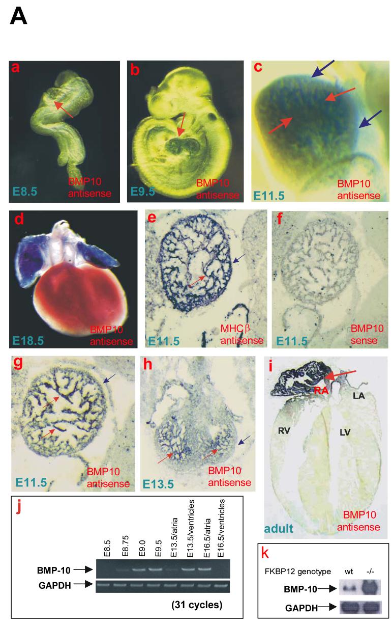
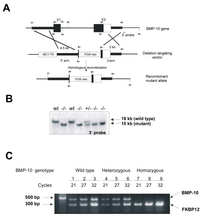
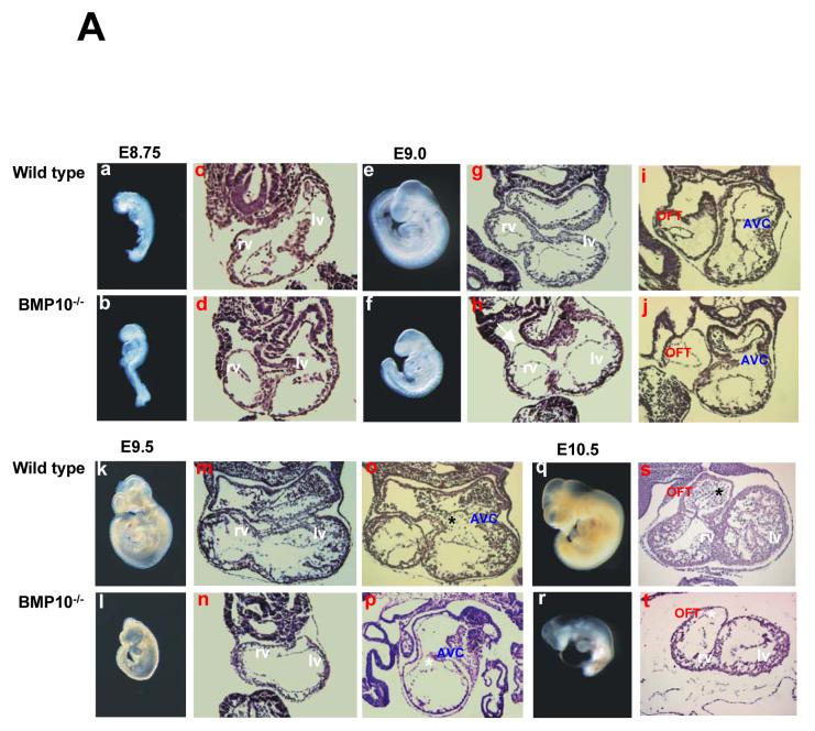
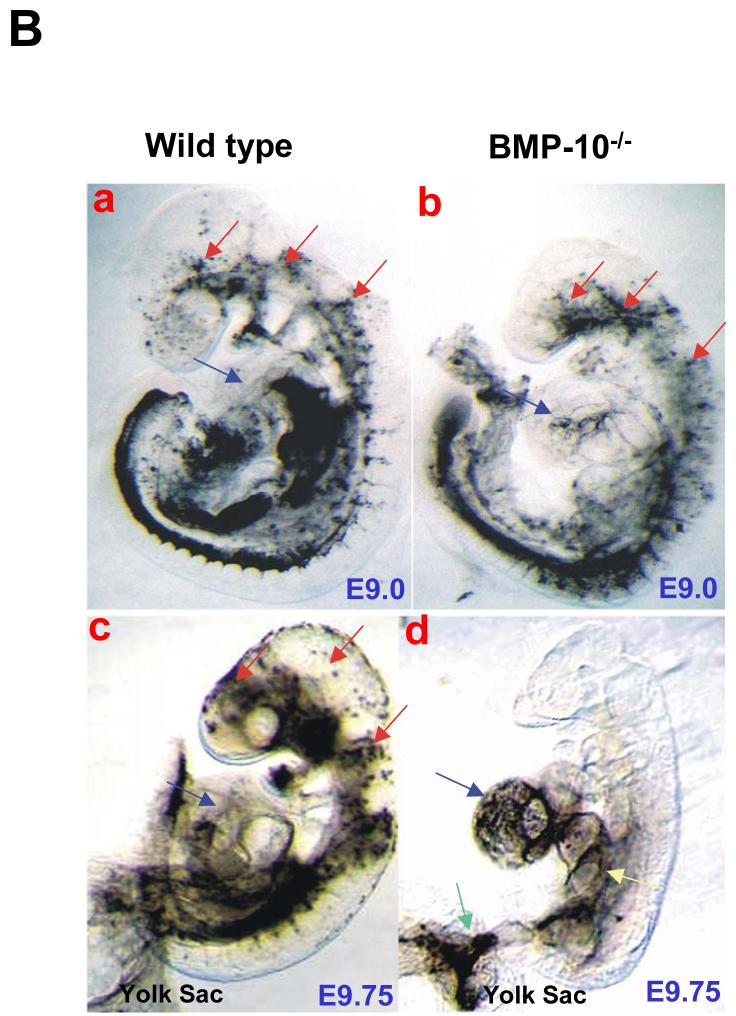
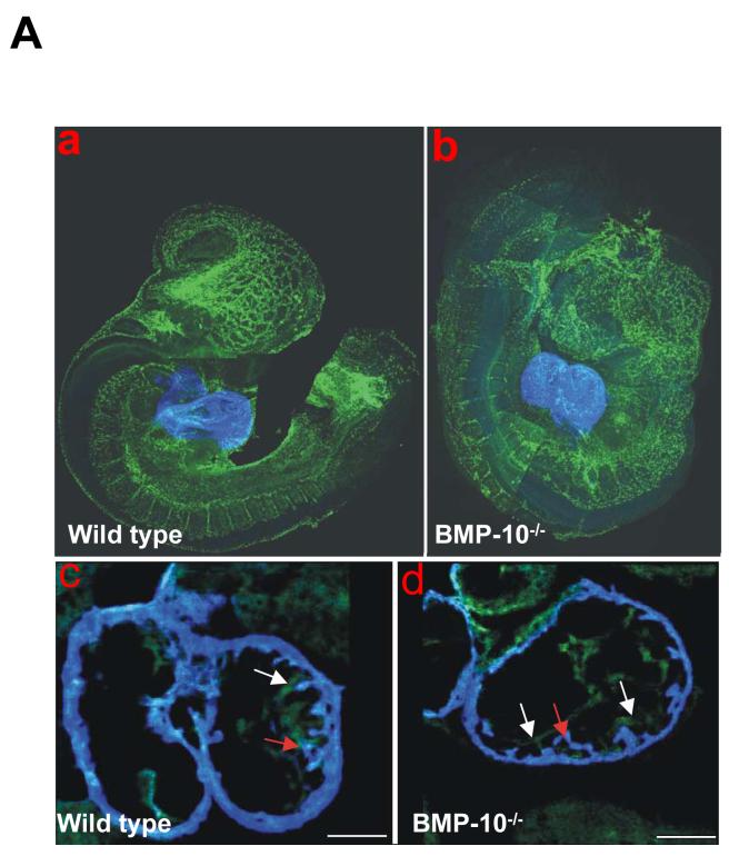
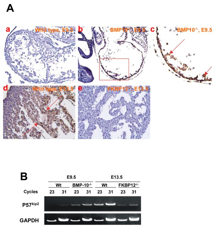
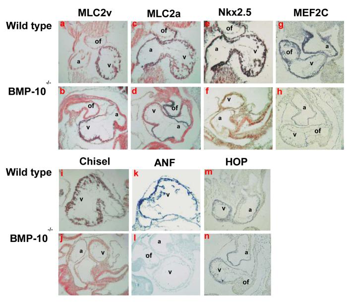
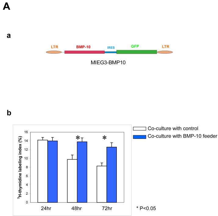
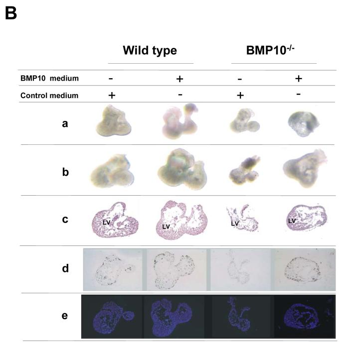
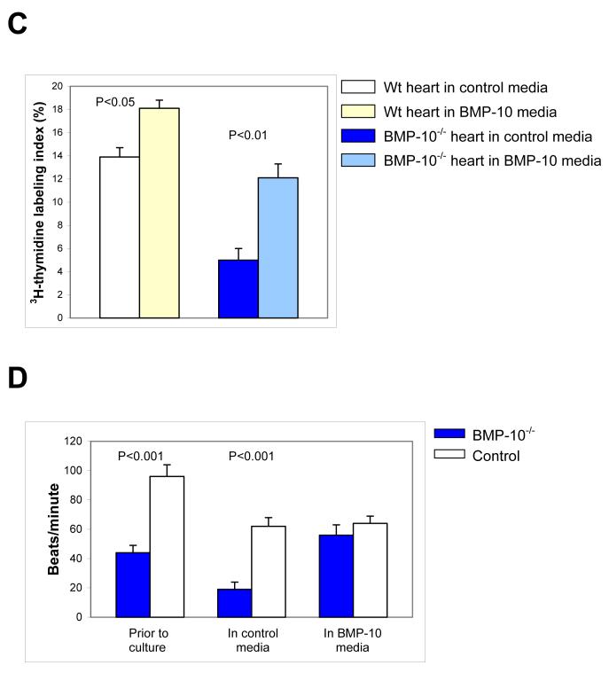
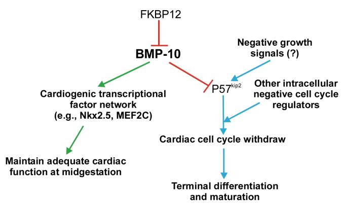
During cardiogenesis, perturbation of a key transition at mid-gestation from cardiac patterning to cardiac growth and chamber maturation often leads to diverse types of congenital heart disease, such as ventricular septal defect (VSD), myocardium noncompaction, and ventricular hypertrabeculation. This transition, which occurs at embryonic day (E) 9.0-9.5 in murine embryos and E24-28 in human embryos, is crucial for the developing heart to maintain normal cardiac growth and function in response to an increasing hemodynamic load. Although, ventricular trabeculation and compaction are key morphogenetic events associated with this transition, the molecular and cellular mechanisms are currently unclear. Initially, cardiac restricted cytokine bone morphogenetic protein 10 (BMP10) was identified as being upregulated in hypertrabeculated hearts from mutant embryos deficient in FK506 binding protein 12 (FKBP12). To determine the biological function of BMP10 during cardiac development, we generated BMP10-deficient mice. Here we describe an essential role of BMP10 in regulating cardiac growth and chamber maturation. BMP10 null mice display ectopic and elevated expression of p57(kip2) and a dramatic reduction in proliferative activity in cardiomyocytes at E9.0-E9.5. BMP10 is also required for maintaining normal expression levels of several key cardiogenic factors (e.g. NKX2.5 and MEF2C) in the developing myocardium at mid-gestation. Furthermore, BMP10-conditioned medium is able to rescue BMP10-deficient hearts in culture. Our data suggest an important pathway that involves a genetic interaction between BMP10, cell cycle regulatory proteins and several major cardiac transcription factors in orchestrating this transition in cardiogenesis at mid-gestation. This may provide an underlying mechanism for understanding the pathogenesis of both structural and functional congenital heart defects.


Figures

Figure 1
Figure 1
Expression pattern of BMP-10 during cardiogenesis. (A) In (a-d), using whole-mount in situ hybridization, BMP-10 transcripts were not detected in E8.5 embryonic hearts (a), but were detected in developing ventricles and atria at E9.5 (b), and at E11.5 (c). BMP-10 expression was restricted to trabecular myocardium. Red arrows indicate the heart in (a and b) and trabecular myocardium in (c), and blue arrows indicate the ventricular compact wall. (d) BMP-10 expression was only detected in atria at E18.5. In (e-i), in situ hybridizations were performed on sagittal sections of E11.5, transverse sections of E13.5 embryos, and transverse sections of an adult mouse heart. (e) Myosin heavy chain β (MHCβ) transcripts were detected throughout the myocardium. (f) BMP-10 sense probe was used as a negative control. (g and h) BMP-10 expression in ventricle was restricted to trabecular myocardium, and (i) became restricted to the right atrium in adult. (j) BMP-10 transcripts were detected in the heart as early as E8.75 using RT-PCR. By E16.5, BMP-10 is hardly detectable in ventricles. (k) Using Northern blot to confirm BMP-10 expression in FKBP12-deficient heart (E14.5). BMP-10 transcripts were significantly up-regulated in FKBP12-deficient heart. (B) Multiple-tissue quantitative mRNA level analysis. (a) Quantitative dot blot analysis of evenly loaded 84 mRNA samples from varying adult human tissues (Clontech). Only mRNA samples isolated from whole heart (A4) and right atrium (D4) showed significant BMP-10 mRNA level when compared to negative control samples. (b) Phosphorimage analysis on the relative level of expression of BMP-10 in different regions of the adult heart. The phosphorimaging values were corrected for the mean background consisting of negative controls (yeast total RNA, yeast tRNA, E.coli rRNA, E. Coli DNA) and empty fields.
Figure 1
Figure 1
Expression pattern of BMP-10 during cardiogenesis. (A) In (a-d), using whole-mount in situ hybridization, BMP-10 transcripts were not detected in E8.5 embryonic hearts (a), but were detected in developing ventricles and atria at E9.5 (b), and at E11.5 (c). BMP-10 expression was restricted to trabecular myocardium. Red arrows indicate the heart in (a and b) and trabecular myocardium in (c), and blue arrows indicate the ventricular compact wall. (d) BMP-10 expression was only detected in atria at E18.5. In (e-i), in situ hybridizations were performed on sagittal sections of E11.5, transverse sections of E13.5 embryos, and transverse sections of an adult mouse heart. (e) Myosin heavy chain β (MHCβ) transcripts were detected throughout the myocardium. (f) BMP-10 sense probe was used as a negative control. (g and h) BMP-10 expression in ventricle was restricted to trabecular myocardium, and (i) became restricted to the right atrium in adult. (j) BMP-10 transcripts were detected in the heart as early as E8.75 using RT-PCR. By E16.5, BMP-10 is hardly detectable in ventricles. (k) Using Northern blot to confirm BMP-10 expression in FKBP12-deficient heart (E14.5). BMP-10 transcripts were significantly up-regulated in FKBP12-deficient heart. (B) Multiple-tissue quantitative mRNA level analysis. (a) Quantitative dot blot analysis of evenly loaded 84 mRNA samples from varying adult human tissues (Clontech). Only mRNA samples isolated from whole heart (A4) and right atrium (D4) showed significant BMP-10 mRNA level when compared to negative control samples. (b) Phosphorimage analysis on the relative level of expression of BMP-10 in different regions of the adult heart. The phosphorimaging values were corrected for the mean background consisting of negative controls (yeast total RNA, yeast tRNA, E.coli rRNA, E. Coli DNA) and empty fields.
Figure 2
Figure 2
Generation of BMP-10-deficient mice. Targeting vector to mutate the mouse BMP-10 gene in ES cells (a), Southern blot analysis of genomic DNA (digested with EcoRV and probed with 3′-probe) derived from a single litter of E9.5 embryos after mating of bmp10m1/+ mice (b), and RT-PCR analysis to confirm the inactivation of BMP-10 expression in bmp10m1/bmp10m1 embryos (E9.0) (c). Expression of FKBP12 was used as loading control.
Figure 3
Figure 3
Morphological and histological analysis of BMP-10-deficient embryos and hearts. (A) In a and b, e and f, k and l, q and r, comparison of gross morphology of normal littermate control and BMP-10-deficient embryos from E8.75 to E10.5. (a and b) No apparent abnormality was detected at E8.75. (e and f) Some BMP10-deficient embryos were slightly growth retarded at E9.0. (k and l) Severe growth retardation was seen in BMP-10-deficient embryo at E9.5, however, mutants had an identical number of somite pairs and normal allantoic connection when compared to littermate controls. Over 50% of BMP-10-deficient embryos have severe edema and expanded pericardiac sacs, suggesting poor cardiac function in these mutants. (q and r) BMP-10-deficient embryos were dead by E10.5. In c and d, g and h, i and j, m and n, o and p, and s and t, comparison of histological sections of normal control and BMP-10-deficient hearts from E8.75 to E10.5 embryos stained with haematoxylin and eosin. (d, h and j) At E8.75-E9.0, BMP-10-deficient embryos had normal rightward looped heart and primitive ventricular chambers, suggesting that BMP-10 is not required for the early phases of cardiogenesis. Also, the size of the heart in BMP-10-deficient embryos was grossly normal compared to littermate control, but exhibited some thinned myocardium (white arrow). Acellular endocardial cushions were formed in both the outflow track (OFT) and atrial-ventricular canal (AVC). (n, p and t) Compared with wild-type normal heart at E9.5-E10.5, BMP-10-deficient hearts were growth retarded, had hypoplastic walls, and failed to develop normal ventricular trabeculae and endocardial cushions. While endocardial cushions in OFT and AVC of wild type control hearts have begun to be seeded after epithelialmesenchymal transformation of adjacent endocardium (black asterisks), acellular endocardial cushions were remained in BMP-10-deficient hearts (white asterisks). (B) Ink-injection was used to visualize the cardiac contractile function and blood flow in E9.0 and E9.5-E9.75 embryos. (a and c) Ink injected in the primitive left ventricle was efficiently pumped throughout entire cardiovascular system in control embryos. (b) At E9.0, the circulation was established in BMP-10-deficient embryos, however, not as efficiently as littermate controls, which might reflect the weaker/slower heart rate in BMP-10 mutants. (d) At E9.5-9.75, ink remained in the BMP-10-deficient ventricles, suggesting a poor cardiac function. Note that the ink within the BMP-10-deficient heart has diffused in a retrograde direction into the sinus venous (yellow arrow) and yolk sac (green arrow) due to lack of adequate cardiac contraction and circulation. Red arrows indicate circulated ink around embryonic head region in a control embryo. Blue arrows indicate hearts. The posterior portions of the embryos were removed to help visualize the hearts (c and d).
Figure 3
Figure 3
Morphological and histological analysis of BMP-10-deficient embryos and hearts. (A) In a and b, e and f, k and l, q and r, comparison of gross morphology of normal littermate control and BMP-10-deficient embryos from E8.75 to E10.5. (a and b) No apparent abnormality was detected at E8.75. (e and f) Some BMP10-deficient embryos were slightly growth retarded at E9.0. (k and l) Severe growth retardation was seen in BMP-10-deficient embryo at E9.5, however, mutants had an identical number of somite pairs and normal allantoic connection when compared to littermate controls. Over 50% of BMP-10-deficient embryos have severe edema and expanded pericardiac sacs, suggesting poor cardiac function in these mutants. (q and r) BMP-10-deficient embryos were dead by E10.5. In c and d, g and h, i and j, m and n, o and p, and s and t, comparison of histological sections of normal control and BMP-10-deficient hearts from E8.75 to E10.5 embryos stained with haematoxylin and eosin. (d, h and j) At E8.75-E9.0, BMP-10-deficient embryos had normal rightward looped heart and primitive ventricular chambers, suggesting that BMP-10 is not required for the early phases of cardiogenesis. Also, the size of the heart in BMP-10-deficient embryos was grossly normal compared to littermate control, but exhibited some thinned myocardium (white arrow). Acellular endocardial cushions were formed in both the outflow track (OFT) and atrial-ventricular canal (AVC). (n, p and t) Compared with wild-type normal heart at E9.5-E10.5, BMP-10-deficient hearts were growth retarded, had hypoplastic walls, and failed to develop normal ventricular trabeculae and endocardial cushions. While endocardial cushions in OFT and AVC of wild type control hearts have begun to be seeded after epithelialmesenchymal transformation of adjacent endocardium (black asterisks), acellular endocardial cushions were remained in BMP-10-deficient hearts (white asterisks). (B) Ink-injection was used to visualize the cardiac contractile function and blood flow in E9.0 and E9.5-E9.75 embryos. (a and c) Ink injected in the primitive left ventricle was efficiently pumped throughout entire cardiovascular system in control embryos. (b) At E9.0, the circulation was established in BMP-10-deficient embryos, however, not as efficiently as littermate controls, which might reflect the weaker/slower heart rate in BMP-10 mutants. (d) At E9.5-9.75, ink remained in the BMP-10-deficient ventricles, suggesting a poor cardiac function. Note that the ink within the BMP-10-deficient heart has diffused in a retrograde direction into the sinus venous (yellow arrow) and yolk sac (green arrow) due to lack of adequate cardiac contraction and circulation. Red arrows indicate circulated ink around embryonic head region in a control embryo. Blue arrows indicate hearts. The posterior portions of the embryos were removed to help visualize the hearts (c and d).
Figure 4
Figure 4
Whole mount immunostaining and confocal microscopic analysis of control (a and c) and BMP-10-deficient embryos (b and d) at E9.25. Fluorescence conjugated anti-Flk-1 monoclonal antibody stains the endothelial cells (green) in the developing vasculature and endocardium of the developing heart, while anti-myosin heavy chain monoclonal antibody MF-20 stains the myocardium (blue). In a and b, comparison of endothelial development in both control and BMP-10-deficient embryos at E9.25. Endothelial development was not affected in BMP-10-deficient embryos. In c and d, comparison of endocardium and myocardium development in control and BMP-10-deficient ventricles. The BMP-10-deficient heart displayed a much thinner ventricular wall compared to control heart, however, the endocardium was in normal proximity to the myocardium. Some primitive trabeculae were formed in the BMP-10-deficient ventricles at this age. Red arrows point to the primitive trabecular myocardium, while white arrows indicate the endocardium.
Figure 5
Figure 5
Distribution and expression of p57kip2 in BMP-10-deficient and FKBP12-deficient hearts using immunohistochemistry staining. (A) At E9.5, p57kip2 expression was undetectable in wild type heart (a), but was ectopically present in the BMP-10-deficient heart (b and c) under identical staining conditions. The expression of p57kip2 was abundant in E13.5 ventricular trabecular myocardium of wild type heart (d), but was significantly down-regulated in the E13.5 FKBP12-deficient trabecular myocardium (e). Arrows indicate areas of positive staining. (B) Using RT-PCR to confirm the mRNA level of p57kip2 in BMP-10-deficient hearts (E9.5) and FKBP12-deficient hearts (E13.5). The expression of p57kip2 was up-regulated in BMP-10-deficient hearts and down-regulated in FKBP12-deficient hearts.
Figure 6
Figure 6
Analysis of cardiac markers in BMP-10-deficient hearts. (A) Using in situ hybridization to analyze the expression of cardiac markers at E9.5. The cardiac chamber markers MLC2v (a and b) and MLC2a (c and d) were not altered in the BMP-10-deficient hearts, suggesting that cardiac patterning and chamber specification are normal in the BMP-10-deficient heart. Cardiogenic transcription factors Nkx2.5 (e and f) and MEF2C (g and h) were significantly down-regulated at E9.5 in the BMP-10-deficient hearts. The expression of Chisel (i and j) and ANF (k and l) was also reduced in the BMP-10-deficient heart, while the expression of HOP (m and n) remained at a similar level in the BMP-10-deficient heart when compared to the control heart. a: atrium; v: ventricle; of: outflow tract.
Figure 7
Figure 7
The effect of BMP-10 to cultured cardiomyocytes and hearts. (A) Generation of BMP-10 expressing NIH 3T3 cell lines and using cardiomyocyte co-culture assay to determine the biological activity of BMP-10. (a) Schematic diagram of BMP-10 retroviral vector used to express BMP-10. (b) Co-culture of embryonic cardiomyocytes with BMP-10 feeder cells (NIH3T3/BMP-10) and control feeder cells (NIH3T3/EGFP). The proliferative activity of cardiomyocytes in culture after 24, 48 and 72 hours were determined by using 3H-thymidine labeling index of PAS positive cells as described in Methods. BMP-10 feeders were able to maintain higher proliferative activity of cardiomyocytes compared to control feeder cells. (B) In vitro culture of embryonic hearts in BMP-10 conditioned and control media. Embryonic hearts were isolated from E9.0-E9.25 embryos harvested from bmp10m1/+ matings. Each heart was photographed before (a) and after (b) 24 hours of culture. (c), haematoxylin and eosin stained histological sections of cultured hearts. (d) 3H-thymidine labeling was used to determine the proliferative activity of cultured hearts. Autoradiographs of 3H-thymidine labeled hearts. (e) Hoechst staining to show nuclei. The images of each column were from the same heart. (C) 3H-thymidine labeling index of cultured embryonic hearts. (D) Heart rates of BMP-10-deficient and control hearts prior to culture, and following culture in control and BMP-10 conditioned media. BMP-10 conditioned media is able to rescue the heart rates of BMP-10-deficient embryos.
Figure 7
Figure 7
The effect of BMP-10 to cultured cardiomyocytes and hearts. (A) Generation of BMP-10 expressing NIH 3T3 cell lines and using cardiomyocyte co-culture assay to determine the biological activity of BMP-10. (a) Schematic diagram of BMP-10 retroviral vector used to express BMP-10. (b) Co-culture of embryonic cardiomyocytes with BMP-10 feeder cells (NIH3T3/BMP-10) and control feeder cells (NIH3T3/EGFP). The proliferative activity of cardiomyocytes in culture after 24, 48 and 72 hours were determined by using 3H-thymidine labeling index of PAS positive cells as described in Methods. BMP-10 feeders were able to maintain higher proliferative activity of cardiomyocytes compared to control feeder cells. (B) In vitro culture of embryonic hearts in BMP-10 conditioned and control media. Embryonic hearts were isolated from E9.0-E9.25 embryos harvested from bmp10m1/+ matings. Each heart was photographed before (a) and after (b) 24 hours of culture. (c), haematoxylin and eosin stained histological sections of cultured hearts. (d) 3H-thymidine labeling was used to determine the proliferative activity of cultured hearts. Autoradiographs of 3H-thymidine labeled hearts. (e) Hoechst staining to show nuclei. The images of each column were from the same heart. (C) 3H-thymidine labeling index of cultured embryonic hearts. (D) Heart rates of BMP-10-deficient and control hearts prior to culture, and following culture in control and BMP-10 conditioned media. BMP-10 conditioned media is able to rescue the heart rates of BMP-10-deficient embryos.
Figure 8
Figure 8
BMP-10 conditioned medium restores normal genetic program in BMP-10-deficient hearts. (A) Whole mount in situ hybridization of Nkx2.5 expression in cultured embryonic hearts. Embryonic hearts were isolated at E9.0 and cultured in BMP-10 conditioned media over night. Prior to the culture, the BMP-10-deficient heart (n=3) (a) had a significantly lower Nkx2.5 expression than the littermate control heart (3) (b). After culture, the BMP-10-deficient heart (n=4) (c) had restored Nkx2.5 expression. (d) Littermate control heart (n=3) cultured in BMP-10 conditioned medium. (B) Immunohistological staining of p57kip2 expression in cultured embryonic hearts. Embryonic hearts were isolated at E9.0 and cultured in BMP-10 conditioned media or control medium over night. (a) wild type heart cultured in control medium (n=6). (b) BMP-10-deficient heart cultured in control medium (n=4). (c) BMP-10-deficient heart cultured in BMP-10 conditioned medium (n=4). The expression of p57kip2 in mutant hearts cultured in BMP-10 conditioned medium was significantly down-regulated when compared to the mutant hearts cultured in control medium.
Figure 8
Figure 8
BMP-10 conditioned medium restores normal genetic program in BMP-10-deficient hearts. (A) Whole mount in situ hybridization of Nkx2.5 expression in cultured embryonic hearts. Embryonic hearts were isolated at E9.0 and cultured in BMP-10 conditioned media over night. Prior to the culture, the BMP-10-deficient heart (n=3) (a) had a significantly lower Nkx2.5 expression than the littermate control heart (3) (b). After culture, the BMP-10-deficient heart (n=4) (c) had restored Nkx2.5 expression. (d) Littermate control heart (n=3) cultured in BMP-10 conditioned medium. (B) Immunohistological staining of p57kip2 expression in cultured embryonic hearts. Embryonic hearts were isolated at E9.0 and cultured in BMP-10 conditioned media or control medium over night. (a) wild type heart cultured in control medium (n=6). (b) BMP-10-deficient heart cultured in control medium (n=4). (c) BMP-10-deficient heart cultured in BMP-10 conditioned medium (n=4). The expression of p57kip2 in mutant hearts cultured in BMP-10 conditioned medium was significantly down-regulated when compared to the mutant hearts cultured in control medium.
Figure 9
Figure 9
A model for the modulation of cardiac growth and function by BMP-10 during midgestation. FKBP12 negatively regulates BMP-10 possibly via its interaction to type I receptor for BMP-10. BMP-10 has double biological activities: 1) prevents the premature activation and/or antagonizes the activity of negative cell cycle regulators such as p57kip2; 2) maintains cardiac function by regulating level of expression of several key cardiogenic transcriptional factors during midgestation.

References

    1. Andree B, Duprez D, Vorbusch B, Arnold HH, Brand T. BMP-2 induces ectopic expression of cardiac lineage markers and interferes with somite formation in chicken embryos. Mech Dev. 1998;70:119–131. - PubMed
    1. Bao S, Shen X, Shen K, Liu Y, Wang XF. The mammalian Rad24 homologous to yeast Saccharomyces cerevisiae Rad24 and Schizosaccharomyces pombe Rad17 is involved in DNA damage checkpoint. Cell Growth Differ. 1998;9:961–967. - PubMed
    1. Barron M, Gao Min., Lough J. Requirement for BMP and FGF signaling during cardiogenic induction in non-precardiac mesoderm is specific, transient, and cooperative. Dev. Mech. 2000;218:383–393. - PubMed
    1. Black BL, Olson EN. Control of cardiac development by the MEF2 family of transcription factors. In: Harvey RP, Rosenthal N, editors. Heart Development. Academia press; 1999. pp. 131–142.
    1. Chen F, Kook H, Milewski Rita., Gitler AD, Lu MM, Nazarian R, Schnepp R, Jen K, Biben C, Runke G, Mackay JP, Novotny J, Schwatz RJ, Harvey RP, Mullins MC, Epstein JA. Hop is an unusual homeobox gene that modulates cardiac development. Cell. 2002;110:713–723. - PubMed

Publication types

MeSH terms