. 2004 Apr 20;101(16):6068-73.
doi: 10.1073/pnas.0401393101. Epub 2004 Apr 8.

Transcriptional termination by RNA polymerase I requires the small subunit Rpa12p

Affiliations

Transcriptional termination by RNA polymerase I requires the small subunit Rpa12p

Elizabeth M Prescott et al. Proc Natl Acad Sci U S A. .

Abstract

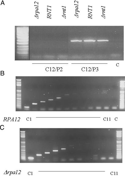
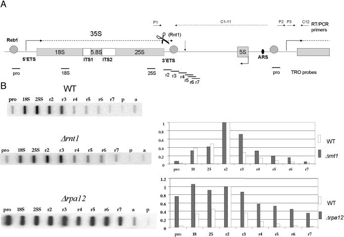
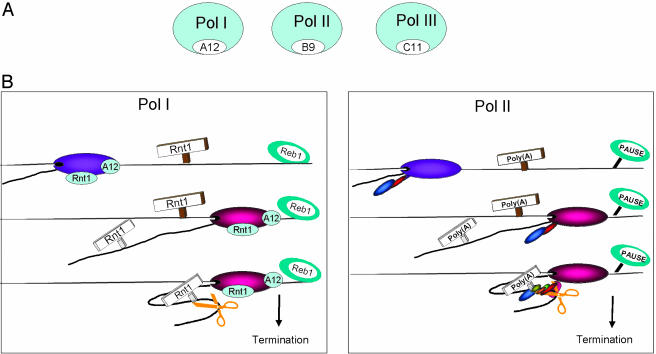
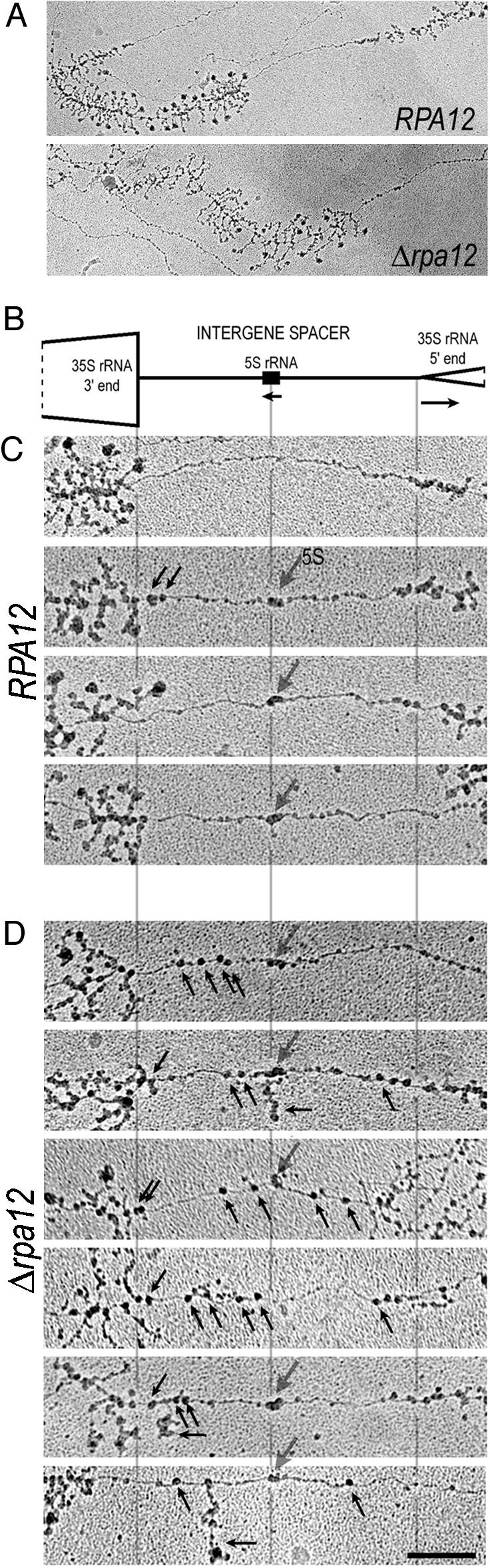
We identify Rpa12p of RNA polymerase I (Pol I) as a termination factor. Combined analyses using transcription run-on, electron microscopy-visualized chromatin spreading and RT-PCR have been applied to the rRNA-encoding genes of Saccharomyces cerevisiae. These confirm that Pol I termination occurs close to the Reb1p-dependent terminator in wild-type strains. However, deletion mutants for the 3' end-processing enzyme Rnt1p or the Rpa12p subunit of Pol I both show Pol I transcription in the spacer. For Deltarpa12, these spacer polymerases are devoid of nascent transcripts, suggesting they are immediately degraded. The homology of Rpa12p to the small subunit Rpb9p of Pol II and Rpc11p of Pol III, both implicated in transcriptional termination, points to a common termination mechanism for all three classes of RNA polymerase.


Figures

Fig. 3.
Fig. 3.
RT-PCR analysis of steady-state rRNA generated in wild-type, Δrnt1, and Δrpa12 strains. (A) RT-PCR across the Pol I transcription start site reveals no detectable steady-state transcription reading into the Pol I promoter region in any strain tested. RT and PCR primers used are indicated, as are their positions relative to the rDNA repeat (Fig. 1 A). PCR products are resolved by agarose gel electrophoresis. (B and C) RT-PCR analysis beyond the normal Reb1p terminator is shown for isogenic wild-type and Δrpa12 strains. Faint signals are detected up to cDNA primer C5, which maps to rDNA nucleotides 7598–7618, but the same profile is found with both strains. cDNAs were generated with primers C1–C11 and amplified by using primer P1.
Fig. 1.
Fig. 1.
TRO analysis of wild-type, Δrnt1, and Δrpa12 strains of S. cerevisiae across the rDNA repeat unit. (A) Diagram indicating the arrangement and location of rRNA sequences within the rDNA repeat unit. ETS and ITS denote external and internal transcribed spacers. The extent of the Pol I pre-rRNA transcript is indicated (dashed line). The positions of terminator and promoter-bound pReb1p are indicated as well as the Rnt1p cleavage site at the end of 35S pre-rRNA. The position of the failsafe terminator in the spacer is indicated by a vertical arrow and the autonomous replication sequence (ARS) by a black oval. The positions of M13 probes are denoted by horizontal bars below the gene map, whereas RT-PCR primers are indicated by horizontal arrows above the gene map (see Materials and Methods for details). (B) TRO data (hybridization signals to slot blots of single-strand DNA M13 phage probes) and their quantitative analysis for the Δrnt1 and Δrpa12 as compared to their isogenic wild-type strains. M13 probe a detects actin gene transcription by Pol II. Quantitation is fixed with all signals relative to probe r2, set as 1. All quantitations are based on multiple repeats of TROs and are corrected for background hybridization (probe p) and U content. The patterns obtained were found to be reproducible within a 10% range. The examples shown are representative images from the whole data set.
Fig. 4.
Fig. 4.
Homologous mechanisms of transcriptional termination for Pol I and II. (A) Homologous small subunits (white shading) in all three polymerase classes. (B) Model showing marked similarity in the mechanisms of Pol I and II termination. Polymerases before the terminal cleavage site are colored purple and after, red, to indicate possible conformational change. The Pol II CTD tail with associated 3′ end processing factors is indicated. Both Pol I and II nascent transcripts are predicted to fold around and interact with paused polymerase. The associated 3′ processing factors will then mediate 3′ end cleavage (orange scissors). This process is facilitated by polymerase pausing, which in turn will induce transcriptional termination. For Pol I, this depends on Reb1p and may require interactions with the Pol I subunit, Rpa12p.
Fig. 2.
Fig. 2.
EM visualization reveals polymerases in the intergenic spacers between rRNA genes of Δrpa12 but not in the spacers in the wild-type strain. (A) Representative 35S rRNA gene/spacer units from the control RPA12 and Δrpa12 strains. The intergenic spacers from these examples are shown at a higher magnification in C Top below (for wild type) and D Top below (for Δrpa12). Clusters of rRNA genes are readily identifiable in yeast chromatin spreads by virtue of their length, tandem repetition, and characteristic terminal knobs on the transcripts. (B) Schematic diagram of the yeast intergenic spacer, drawn to scale. (C) Four examples of intergenic spacers from the control RPA12 strain, aligned with the schematic in B. These spacers are typically free of polymerases, with two general exceptions. First, polymerases with no or very short transcripts are occasionally seen very near the 3′ end of the 35S rRNA gene (black downward-pointing arrows), presumably representing Rnt1 cleavage of nascent 35S rRNA. Second, a characteristic peanutshaped structure (larger gray downward arrow) is often seen at the position of the 5S rRNA gene, presumably representing a few Pol III molecules and associated transcription factors. Nucleosomes are also sometimes seen in the spacers, such as C Bottom. Nucleosomes are smaller and less electron-dense than polymerases. (D) Six examples of intergenic spacers from the mutant Δrpa12 strain, aligned with the schematic in B. Seventy percent of the genes in this strain exhibited spacer polymerases in addition to the two expected types, as explained in C (again shown with downward arrows). Most of the spacer polymerases in the mutant strain have either no or very short transcripts (black upward-pointing arrows), although some have a long transcript that originated in the 35S gene (horizontal arrows). (Bar = 0.2 μm.)

References

    1. Reeder, R. H. (1990) Trends Genet. 6, 390-395. - PubMed
    1. Orphanides, G., Lagrange, T. & Reinberg, D. (1996) Genes Dev. 10, 2657-2683. - PubMed
    1. Geiduschek, E. P. & Kassavetis, G. A. (2001) J. Mol. Biol. 310, 1-26. - PubMed
    1. White, R. J. (2001) Gene Transcription: Mechanisms and Control (Blackwell, Oxford).
    1. McKnight, S. L. (1996) Genes Dev. 10, 367-381. - PubMed

Publication types

MeSH terms