. 2004 Apr;3(2):348-58.
doi: 10.1128/EC.3.2.348-358.2004.

Role of a mitogen-activated protein kinase pathway during conidial germination and hyphal fusion in Neurospora crassa

Affiliations

Role of a mitogen-activated protein kinase pathway during conidial germination and hyphal fusion in Neurospora crassa

Amita Pandey et al. Eukaryot Cell. 2004 Apr.

Abstract

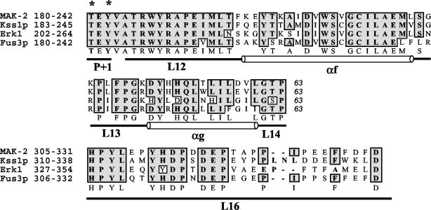
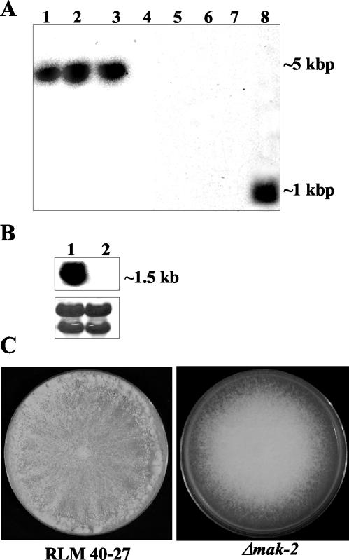
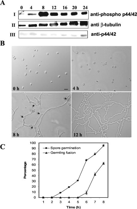
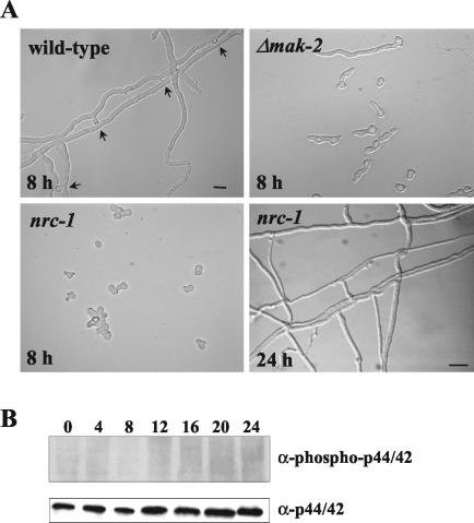
Mitogen-activated protein (MAP) kinase signaling pathways are ubiquitous and evolutionarily conserved in eukaryotic organisms. MAP kinase pathways are composed of a MAP kinase, a MAP kinase kinase, and a MAP kinase kinase kinase; activation is regulated by sequential phosphorylation. Components of three MAP kinase pathways have been identified by genome sequence analysis in the filamentous fungus Neurospora crassa. One of the predicted MAP kinases in N. crassa, MAK-2, shows similarity to Fus3p and Kss1p of Saccharomyces cerevisiae, which are involved in sexual reproduction and filamentation, respectively. In this study, we show that an N. crassa mutant disrupted in mak-2 exhibits a pleiotropic phenotype: derepressed conidiation, shortened aerial hyphae, lack of vegetative hyphal fusion, female sterility, and autonomous ascospore lethality. We assessed the phosphorylation of MAK-2 during conidial germination and early colony development. Peak levels of MAK-2 phosphorylation were most closely associated with germ tube elongation, branching, and hyphal fusion events between conidial germlings. A MAP kinase kinase kinase (NRC-1) is the predicted product of N. crassa nrc-1 locus and is a homologue of STE11 in S. cerevisiae. An nrc-1 mutant shares many of the same phenotypic traits as the mak-2 mutant and, in particular, is a hyphal fusion mutant. We show that MAK-2 phosphorylation during early colony development is dependent upon the presence of NRC-1 and postulate that phosphorylation of MAK-2 is required for hyphal fusion events that occur during conidial germination.


Figures

FIG. 1.
FIG. 1.
Alignment of MAK-2, Kss1p, Erk1, and Fus3p. All four of the MAP kinases are aligned by Clustal W with identity-based alignment. The regions presented are the catalytic core (63 amino acids, P+1 to L14) and the substrate interaction and docking domain (L16). All four of the MAP kinases are activated by phosphorylation at T*EY* in the P+1 loop in the catalytic domain. The phosphorylation lip starts from amino acids Asp-Phe-Gly (DFG) in subdomain VII and ends at TEY in subdomain VIII. Structural domains (L, loop; α, α-helix) are designated according established nomenclature (1). National Center for Biotechnology Information accession numbers are as follows: mak-2, AF348490; FUS3, Z35777; KSS1, Z72825; Erk1, P27361. Asterisks indicate phosphorylated residues.
FIG. 2.
FIG. 2.
Analysis of wild-type and Δmak-2 strains. (A) Southern analysis of genomic DNA from Δmak-2 strains and RLM 40-27 (wild-type strain). Genomic DNA was digested with EcoRV and probed with a hygromycin phosphotransferase gene fragment (lanes 1 to 4) or a mak-2 PCR fragment (lanes 5 to 8). Δmak-2 progeny were APJ-2 (lanes 1 and 5), APJ-1 (lanes 2 and 6), PB-1 (lanes 3 and 7), and RLM 40-27 (lanes 4 and 8). (B) Northern analysis of mak-2 transcription in RLM 40-27 versus Δmak-2 strain PB-1. Two-day-old mycelia were used for RNA extraction. A mak-2 PCR fragment was used as a probe (see Materials and Methods). Lane 1, RLM 40-27; lane 2, PB-1. The same blot was stained with methylene blue as an RNA loading control (lower panel). The rRNA bands are shown. (C) Growth characteristics and morphology of a Δmak-2 mutant compared to wild-type (RLM 40-27). Conidia from RLM 40-27 and PB-1 were inoculated onto plates containing Vogel's (57) minimal medium and assessed for growth and conidiation. Δmak-2 mutants grow slower than the wild type (2.2 versus 7 cm/day) and have derepressed conidiation.
FIG. 3.
FIG. 3.
The Δmak-2 mutant fails to undergo hyphal fusion. Hyphae of wild-type RLM 40-27 and Δmak-2 strains were stained with FM4-64 and imaged by confocal microscopy. Bars, 20 μm. (A) Morphologies of wild-type (RLM 40-27) and Δmak-2 PB-1 strains in the colony periphery. Hyphae do not undergo fusion in this region of the colony in either strain. The Δmak-2 mutant PB-1 shows a branching frequency defect and more meandering hyphae. (B) Morphologies of wild-type (RLM 40-27) and Δmak-2 PB-1 strains in the colony interior. Hyphae of the wild type have undergone many fusion events (each indicated by a white star). In the Δmak-2 mutant, hyphal fusions are absent, although hyphae frequently make physical contact with each other (indicated by a dagger). This figure was provided by David Jacobson.
FIG. 4.
FIG. 4.
The Δmak-2 mutant lacks a protein that is recognized by anti-p44/42 and anti-phospho p44/42 antibodies. (A) Total protein (30 μg) was extracted from wild-type strains (RLM 40-27 and Xa-2), Δmak-2 (PB-1), Δmak-2 progeny (APJ-1, APJ-3, and APJ-4), and nrc-1 and separated on an SDS-10% polyacrylamide gel electrophoresis gel. The blot was probed with anti-p44/42 (α-p44/42) antibodies (PhosphoPlus antibody kit; Cell Signaling Technology). Lane 1, nrc-1; lane 2, APJ-4; lane 3, APJ-3; lane 4, APJ-1; lane 5, RLM 40-27; lane 6, Xa-2; lane 7, PB-1. The lower panel shows blots probed with anti-β-tubulin antibodies for protein loading controls. (B) The introduction of mak-2 into Δmak-2 progeny APJ-1 restores the production of a protein that is recognized by anti-p44/42 antibodies. Total protein (30 μg) was extracted from APJ-1 transformants containing pOKEmak-2 (4C, 6C, and 7C), the wild type (RLM 47-20), and PB-1 Δmak-2 mycelia and separated by SDS-10% polyacrylamide gel electrophoresis followed by transfer onto a nitrocellulose membrane. Anti-p44/42 antibody (Cell Signaling Technology) was used for probing the blot. Lane 1, RLM 40-27; lane 2, Δmak-2 (PB-1); lane 3, APJ-1 transformant (4C) containing pOKEmak-2; lane 4, APJ-1 transformant (6C) containing pOKEmak-2; lane 5, APJ-1 transformant (7C) containing pOKEmak-2. (C) The Δmak-2 mutants lack a protein that is recognized by anti-phospho p44/42 antibodies. Total protein (30 μg) was extracted from the wild-type strain (RLM 40-27), Δmak-2 progeny (APJ-1, APJ-2, and APJ-4) and nrc-1 grown for 20 h as described in Materials and Methods and separated on an SDS-10% polyacrylamide gel electrophoresis gel. The blot was probed with anti-phospho p44/42 antibodies (PhosphoPlus antibody kit; Cell Signaling Technology). Lane 1, nrc-1; lane 2, APJ-1; lane 3, APJ-4; lane 4, APJ-2; lane 5, RLM 40-27. (D) The APJ-1 transformant (6C) containing mak-2 shows restoration of hyphal fusion. Conidia from the APJ-1 transformant containing pOKEmak-2 (6C) were inoculated onto cellophane layered onto plates containing Vogel's minimal medium. Colonies were assessed for hyphal fusion events after 24 h of growth. Arrows indicate hyphal fusion events. Bar, 10 μm.
FIG. 5.
FIG. 5.
MAK-2 phosphorylation is associated with germ tube elongation and hyphal fusion between germlings. (A) Conidia from a wild-type strain (RLM 40-27) were inoculated onto a cellophane membrane layered on Vogel's minimal medium plates and incubated at 24°C. Total protein (30 μg) isolated at 0, 4, 8, 12, 16, 20, and 24 h postinoculation of conidia was used for Western blot analysis. (I) Protein extracts from the different time points probed with anti-phospho p44/42 antibodies (PhosphoPlus antibody kit; Cell Signaling Technology). (II) The blot in panel I was stripped and reprobed with anti-β-tubulin monoclonal antibodies (clone TU27; BabCo). (III) The blot in panel II was restripped and probed with anti-p44/42 antibodies. The anti-phospho p44/p42 antibodies (Cell Signaling Technology) recognize highly conserved phosphorylated T and Y residues in MAP kinases (residues 202 to 204 in Erk1 and residues 180 to 182 in MAK-2) (Fig. 1). The anti-p44/42 antibodies were raised to a peptide synthesized based on the C-terminal amino acid sequence of Erk1. The phospho-p44/42 antibody gave a stronger signal on Western blots than the anti-p44/42 antibody, presumably because of the highly conserved TEY site in MAK-2. The same experiment was repeated with unstripped blots for anti-p44/42 antibodies, and identical results were obtained. (B) DIC images of conidia and conidial germlings from the wild type (RLM 40-27) at 0, 4, 8, and 12 h postinoculation. Bar, 30 μm. (C) Quantitation of germination and hyphal fusion in conidial germlings in the wild type (RLM 40-27) over the first 8 h postinoculation. Error bars represent standard errors.
FIG. 6.
FIG. 6.
nrc-1 is required for phosphorylation of MAK-2 during conidial germination. (A) DIC image of germinating conidia from wild-type (RLM 40-27), Δmak-2, and nrc-1 strains 8 h postinoculation and from the nrc-1 strain 24 h postinoculation. Arrows indicate fusion events observed in RLM 40-27. Hyphal fusion was not evident in nrc-1, although hyphae frequently made contact or grew over each other (24 h). Bar, 30 μm. (B) Conidia from nrc-1 were inoculated on a cellophane membrane layered onto plates containing Vogel's minimal medium with dextrose at 24°C (nrc-1 is an invertase mutant). Total protein (30 μg) from nrc-1 mycelia was harvested at the 0-, 4-, 8-, 12-, 16-, 20-, and 24-h time points. Protein extracts were separated by SDS-10% polyacrylamide gel electrophoresis and blotted onto nitrocellulose membranes. (Top panel) Blot probed with anti-phospho p44/42 antibodies (PhosphoPlus antibody kit; Cell Signaling Technology). (Bottom panel) Blot from the panel above stripped and probed with anti-p44/42 antibodies. The experiment was repeated three times with identical results.

Similar articles

Cited by

References

    1. Atienza, J. M., M. Suh, I. Xenarios, R. Landgraf, and J. Colicelli. 2000. Human ERK1 induces filamentous growth and cell wall remodeling pathways in Saccharomyces cerevisiae. J. Biol. Chem. 275:20638-20646. - PubMed
    1. Banuett, F. 1998. Signalling in the yeasts: an informational cascade with links to the filamentous fungi. Microbiol. Mol. Biol. Rev. 62:249-274. - PMC - PubMed
    1. Bistis, G. N. 1981. Chemotropic interactions between trichogynes and conidia of opposite mating-type in Neurospora crassa. Mycologia 73:959-975.
    1. Bobrowicz, P., R. Pawlak, A. Correa, D. Bell-Pedersen, and D. J. Ebbole. 2002. The Neurospora crassa pheromone precursor genes are regulated by the mating type locus and the circadian clock. Mol. Microbiol. 45:795-804. - PubMed
    1. Borneman, A. R., M. J. Hynes, and A. Andrianopoulos. 2001. An STE12 homolog from the asexual, dimorphic fungus Penicillium marneffei complements the defect in sexual development of an Aspergillus nidulans steA mutant. Genetics 157:1003-1014. - PMC - PubMed

Publication types

MeSH terms

Substances

LinkOut - more resources