. 2004 Apr;3(2):459-70.
doi: 10.1128/EC.3.2.459-470.2004.

Tfs1p, a member of the PEBP family, inhibits the Ira2p but not the Ira1p Ras GTPase-activating protein in Saccharomyces cerevisiae

Affiliations

Tfs1p, a member of the PEBP family, inhibits the Ira2p but not the Ira1p Ras GTPase-activating protein in Saccharomyces cerevisiae

Hélène Chautard et al. Eukaryot Cell. 2004 Apr.

Abstract

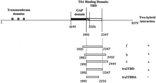
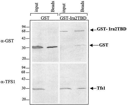
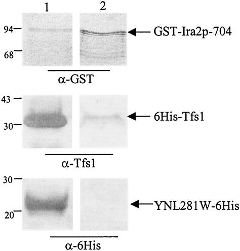
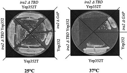
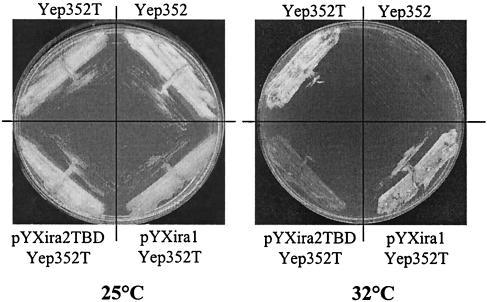
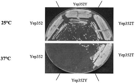
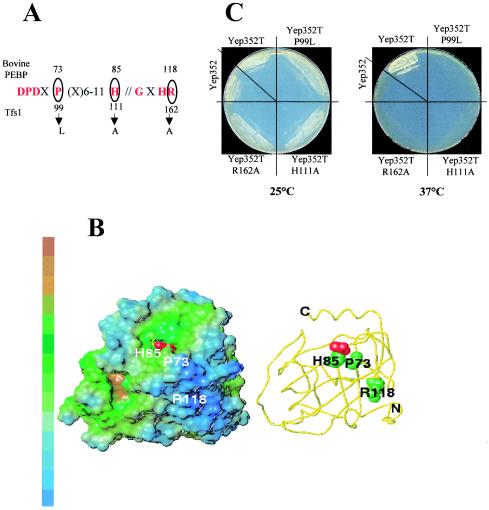
Ras proteins are guanine nucleotide-binding proteins that are highly conserved among eukaryotes. They are involved in signal transduction pathways and are tightly regulated by two sets of antagonistic proteins: GTPase-activating proteins (GAPs) inhibit Ras proteins, whereas guanine exchange factors activate them. In this work, we describe Tfs1p, the first physiological inhibitor of a Ras GAP, Ira2p, in Saccharomyces cerevisiae. TFS1 is a multicopy suppressor of the cdc25-1 mutation in yeast and corresponds to the so-called Ic CPY cytoplasmic inhibitor. Moreover, Tfs1p belongs to the phosphatidylethanolamine-binding protein (PEBP) family, one member of which is RKIP, a kinase and serine protease inhibitor and a metastasis inhibitor in prostate cancer. In this work, the results of (i) a two-hybrid screen of a yeast genomic library, (ii) glutathione S-transferase pulldown experiments, (iii) multicopy suppressor tests of cdc25-1 mutants, and (iv) stress resistance tests to evaluate the activation level of Ras demonstrate that Tfs1p interacts with and inhibits Ira2p. We further show that the conserved ligand-binding pocket of Tfs1-the hallmark of the PEBP family-is important for its inhibitory activity.


Figures

FIG. 1.
FIG. 1.
Ira2p domains and truncated Ira2 protein constructs. The putative membrane-spanning segments are indicated by gray-shaded boxes. The GAP domain is indicated by a filled box; the TBD is indicated by an open box. The overlap between the two domains is hatched. The first and last residues of Ira2p and the limits of the GAP and TBDs are indicated. f, b, and c designate the three IRA2 DNA fragments isolated from positive-testing prey plasmids. + indicates that an interaction was detected by the two-hybrid system; − indicates that no interaction was detected.
FIG. 2.
FIG. 2.
Tfs1p specifically interacts with the GST-Ira2 TBD in vivo. Wild-type yeast cells producing GST alone or GST fused at the N terminus of Ira2 TBD (GST-Ira2TBD) were lysed. An aliquot of these cell extracts (input) was directly analyzed by Western blotting with the antibodies indicated on the left side of the figure. Another aliquot was incubated with glutathione beads, which specifically bind to GST. After several washes, the material bound to the beads was eluted with SDS-PAGE sample buffer (Beads) and analyzed by Western blotting. The molecular-mass markers are indicated (in kilodaltons) on the left side of the figure.
FIG. 3.
FIG. 3.
6His-Tfs1 and GST-Ira2p704 interact in vitro. Purified 6His-Tfs1 or YNL281W-6His (an aliquot of which was analyzed [lane 1] with anti-Tfs1 and anti-six-His antibodies, respectively) was incubated with glutathione beads previously used for affinity purification of GST-Ira2p-704 produced from E. coli (10 μl of extracts was loaded in lane 1 and revealed with anti-GST antibodies). After several washes, the material bound to the beads was eluted with glutathione and analyzed by Western blot analysis with antibodies directed against GST, Tfs1, or six-His (as indicated) (lane 2). The molecular mass markers are indicated (in kilodaltons) on the left side of the figure.
FIG. 4.
FIG. 4.
The TFS1 multicopy suppressor phenotype of cdc25-1 mutants is dependent on the presence of an intact Ira2 TBD. LRB27 cells expressing a wild-type Ira2p (LRB27), Ira2p lacking its 1,435 C-terminal residues (LRB27Δira2GAP), or Ira2 lacking its 847 C-terminal residues (LRB27Δira2TBD) was transformed or not transformed with the following plasmids: Yep352 and Yep352T. Each was then streaked out for single colonies and grown at 25°C or at the restrictive temperature of 37°C on YPD medium.
FIG. 5.
FIG. 5.
The overproduction of Ira2 TBD specifically inhibits the TFS1 multicopy suppressor phenotype of cdc25-1 mutants. LRB27 cells were transformed with the following plasmids: Yep352, Yep352T, pYXIra2TBD, and pYXIra1. They were then streaked out for single colonies and grown at 25°C or at the restrictive temperature of 32°C on YPGal medium to allow Ira2 TBD or Ira1p fragment overproduction.
FIG. 6.
FIG. 6.
YLR179c does not exert a multicopy suppressor function on cdc25-1 mutants. LRB27 cells were transformed with the following plasmids: Yep352, Yep352T, and Yep352Y. They were then streaked out for single colonies at 25°C or at the restrictive temperature of 37°C on YPD medium.
FIG. 7.
FIG. 7.
Physiological effects of TFS1 disruption or overexpression. (A) BY4742 cells disrupted or not disrupted in TFS1 (ΔT and wild type [WT], respectively) were grown to saturation for 2 days, incubated at 37°C for 1 h, and then incubated for various times at 55°C. They were then cooled, and 10-fold serial dilutions were plated on YPD to compare survival. (B) The same experiment was performed with BY4742 cells overproducing or not overproducing Tfs1p (Yep352T or Yep352, respectively).
FIG. 8.
FIG. 8.
Functional importance of conserved residues in Tfs1p. (A) Mutations in the conserved region of Tfs1p. Residues in the conserved regions are indicated in one-letter code. “X” stands for any residue. The circled residues were mutated. The numbers at the top indicate the positions of the corresponding residues in bovine PEBP. The numbers at the bottom indicate the positions of the mutated residues of Tfs1p, and the arrows indicate the mutations. (B) Positions of the mutated residues in the 3D structure of bovine PEBP, showing their locations relative to the putative active site of the bovine PEBP. The residues are numbered as for PEBP. The acetate ligand of the X-ray structure (45) is displayed in a red spacefill representation. Left panel: lipophilic potentials on the surface. The MOLCAD surface (SYBYL software; TRIPOS, St. Louis, Mo.) is colored from blue to brown (indicating increasing hydrophobicity). Right panel (with the same orientation): the PEBP backbone is displayed as a yellow tube; the heavy atoms of the mutated residues P73, H85, and R118 are represented in a spacefill representation. (C) Multicopy suppressor phenotype of Tfs1p mutants. LRB27 cells were transformed with the following plasmids: Yep352, Yep352T, Yep352TP99L, Yep352TH111A, and Yep352TR162A. They were then streaked out for single colonies at 25°C or at the restrictive temperature of 37°C on YPD medium.

Similar articles

Cited by

References

    1. Ahmadian, M. R., P. Stege, K. Scheffzek, and A. Wittinghofer. 1997. Confirmation of the arginine-finger hypothesis for the GAP-stimulated GTP-hydrolysis reaction of Ras. Nat. Struct. Biol. 4:686-689. - PubMed
    1. Banfield, M. J., J. J. Barker, A. C. Perry, and R. L. Brady. 1998. Function from structure? The crystal structure of human phosphatidylethanolamine-binding protein suggests a role in membrane signal transduction. Structure 6:1245-1254. - PubMed
    1. Banfield, M. J., and R. L. Brady. 2000. The structure of Antirrhinum centroradialis protein (CEN) suggests a role as a kinase regulator. J. Mol. Biol. 297:1159-1170. - PubMed
    1. Boguski, M. S., and F. McCormick. 1993. Proteins regulating Ras and its relatives. Nature 366:643-654. - PubMed
    1. Bollag, G., and F. McCormick. 1991. Differential regulation of rasGAP and neurofibromatosis gene product activities. Nature 351:576-579. - PubMed

Publication types

MeSH terms