. 2004 Apr;3(2):483-94.
doi: 10.1128/EC.3.2.483-494.2004.

Cryptosporidium parvum mitochondrial-type HSP70 targets homologous and heterologous mitochondria

Affiliations

Cryptosporidium parvum mitochondrial-type HSP70 targets homologous and heterologous mitochondria

Jan Slapeta et al. Eukaryot Cell. 2004 Apr.

Abstract

A mitochondrial HSP70 gene (Cp-mtHSP70) is described for the apicomplexan Cryptosporidium parvum, an agent of diarrhea in humans and animals. Mitochondrial HSP70 is known to have been acquired from the proto-mitochondrial endosymbiont. The amino acid sequence of Cp-mtHSP70 shares common domains with mitochondrial and proteobacterial homologues, including 34 amino acids of an NH2-terminal mitochondrion-like targeting presequence. Phylogenetic reconstruction places Cp-mtHSP70 within the mitochondrial clade of HSP70 homologues. Using reverse transcription-PCR, Cp-mtHSP70 mRNA was observed in C. parvum intracellular stages cultured in HCT-8 cells. Polyclonal antibodies to Cp-mtHSP70 recognize a approximately 70-kDa protein in Western blot analysis of sporozoite extracts. Both fluorescein- and immunogold-labeled anti-Cp-mtHSP70 localize to a single mitochondrial compartment in close apposition to the nucleus. Furthermore, the NH2-terminal presequence of Cp-mtHSP70 can correctly target green fluorescent protein to the single mitochondrion of the apicomplexan Toxoplasma gondii and the mitochondrial network of the yeast Saccharomyces cerevisiae. When this presequence was truncated, the predicted amphiphilic alpha-helix was shown to be essential for import into the yeast mitochondrion. These data further support the presence of a secondarily reduced relict mitochondrion in C. parvum.


Figures
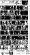
FIG. 1.
FIG. 1.
Multiple protein sequence alignment of cytosolic and mitochondrial HSP70. Conceptually translated full-length amino acid sequences of C. parvum together with S. cerevisiae and P. falciparum were aligned using the program Clustal X and the BLOSUM protein weight matrix. The upper three sequences are mitochondrial; the lower three are cytosolic. Black shading indicates that identical amino acids are conserved, whereas gray indicates conservation of similar amino acids. The threshold for shading was set to 50%. A solid line denotes the predicted Cp-mtHSP70 mitochondrial presequence; a dashed underline denotes the SSC1 yeast mitochondrial presequence (43). The two mitochondrial/proteobacterial sequence signatures motifs, GDAWV and YSPSQI, are denoted with an asterisk above the alignment. The species name and GenBank accession numbers are indicated at the right of the alignment.
FIG. 2.
FIG. 2.
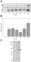
Transcription and expression of C. parvum mtHSP70. (A) RT-PCR analysis of expression during in vitro development in HCT-8 cells and sporozoites (spor.). Polyacrylamide gel signals (Cp-mtHSP70, SSU rRNA) were analyzed using ImageQuantNT (Molecular Dynamics) after 24 cycles. (B) Graph indicating amount of signal after 24 PCR cycles normalized to the amount of SSU rRNA signal detected in each of the infected samples. Three independent experiments were analyzed. (C) Western blot analysis of a crude extract of 107 C. parvum sporozoites. Preimmune sera (lane 1) and immune anti-Cp-mtHSP70 sera (lane 2) are shown. The antisera recognize a single band (arrowhead) of ∼70 kDa in size.
FIG. 3.
FIG. 3.
Phylogenetic tree reconstruction of DnaK and HSP70 homologues. The tree was calculated with the MrBayes program using the JTT model and rooted on Methanothermobacter and Thermoplasma sequences. The numbers on branches are PP MrBayes/bootstrap support ProML, with only values of >0.5/50 shown. GenBank accession numbers for bacterial and archeal DnaK are as follows: Aquifex aeolicus, AAC07071; Borrelia burgdorferi, AAC66887; Chlamydia trachomatis, AAC3683; Ehrlichia sennetsu, AAC27487; Escherichia coli, AAC73125; Francisella tularensis, AAA69561; Haemophilus influenzae, AAC22889; Halobacterium salinarum, AAC41461; Leptospira interrogans, AAC35416; Methanothermobacter thermautotrophicus, CAA14651; Agrobacterium tumefaciens, CAA60592; Mycobacterium avium paratuberculosis, CAA42063; Rhodopseudomonas sp., BAA19796; Rickettsia prowazekii, CAA14651; Sinorhizobium meliloti, AAA64925; Thermoplasma acidophilum, AAC41460. GenBank accession numbers for eukaryote homologues, cytosolic, are as follows: Babesia bovis, AAF14194; Blastocladiella emersonii, AAA65099; Caenorhabditis elegans, AAA28078; Cryptosporidium parvum, AAC25925; Drosophila melanogaster, AAA28625; Giardia lamblia, EAA38588; Leishmania major, P14834; Mus musculus, AAA37864; Pisum sativum, CAA67867; Plasmodium falciparum, NP_704366; Saccharomyces cerevisiae, AAC37398; Schizosaccharomyces pombe, BAA25322; Theileria annulata, A44985; Trichomonas vaginalis, AAB52423; Trypanosoma cruzi, AAA30205. GenBank accession numbers for endoplasmatic reticulum (ER) HSP70s are as follows: Drosophila melanogaster, AAA28626; Giardia lamblia, EAA41481; Plasmodium falciparum, NP_704718; Trypanosoma brucei, AAC37174; Zea mays, AAC49900. GenBank accession numbers for mitochondrial HSP70s are as follows: Arabidopsis thaliana, AAO00750; Caenorhabditis elegans, AAB42371; Cryptosporidium parvum, AAP59793; Drosophila melanogaster, AAA28628; Eimeria tenella, CAA87086; Encephalitozoon cuniculi, CAA10035; Entamoeba histolytica, AAG16651; Giardia intestinalis, BAB84357; Homo sapiens, AAA67526; Leishmania major, P12076; Nosema locustae, AAC47660; Plasmodium falciparum, BAB17688; Saccharomyces cerevisiae, AAA63792; Schizosaccharomyces pombe, AAA35314; Solanum tuberosum, Q08276; Trichomonas vaginalis, AAB09772; Trypanosoma cruzi, AAA30215.
FIG. 4.
FIG. 4.
Presequence of C. parvum mtHSP70. (A) Secondary structure prediction. Line 1 (SSpro8): H, alpha-helix; G, 310-helix; I, pi-helix; E, extended strand; B, beta-bridge; T, turn; S, bend; C, the rest. Line 2 (Psi-Pred): H, helix; E, strand; C, coil. Line 3 (Predator): H, helix; E, extended sheet; _, coil. Line 4: amino acid sequence of the Cp-mtHSP70 NH2 terminus. Eisenberg's hydrophobic moment plot is immediately beneath and aligned with the amino acid sequence above it. The arrow indicates the R-2 motif, xRx↓x(S/x). Numbering denotes amino acid residues. (B) Helical wheel plot of residues 15 to 32 with putative amphiphilic α-helix demonstrating partitioning of charged and hydrophobic amino acids (gray and black circles, respectively). The helical wheel plot was generated with the aid of Java-applet at http://cti.itc.virginia.edu/∼cmg/Demo/wheel/wheelApp.html. (C) Truncated NH2-terminal amino acid sequences of Cp-mtHSP70 cloned into the E. coli/yeast shuttle vector pYX122-GFP, including the yeast SSC1 control (sequence in italic). Numbering above denotes amino acid residues. Constructs are named on the right. (D) Graph of the import efficiency of different GFP constructs based on Western blotting of subcellular fractions. Import efficiency is the ratio between mitochondrially imported GFP and total GFP (mitochondrial and cytosolic) relative to the control construct SSC1. Results are calculated from three independent experiments. Numbering of the constructs corresponds to that in panel 4C.
FIG. 5.
FIG. 5.
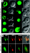
Targeting of C. parvum mtHSP70 presequence to heterologous mitochondria. (A) Targeting of the yeast S. cerevisiae using the Cp-mtHSP70 presequence at the NH2 terminus of GFP vectors (see Fig. 4C). Each panel shows paired GFP and differential interference contrast (DIC) images. Full C. parvum presequence (Cp), negative control lacking the presequence (targeting the cytosol; cGFP), positive control—the yeast mtHSP70 homologue SSC1 presequence, targeting the yeast mitochondrial network (Sc), and the C. parvum truncated forms, Δ1-10, Δ1-20, Δ1-30, Δ10-20, Δ10-30, and Δ10-40, indicating the regions of the presequence excised, are shown. Bar, 5 μm. (B) Targeting of GFP using the Cp-mtHSP70 presequence at the NH2 terminus of GFP (upper panels, GRA1-[pre]-GFP) or no-presequence control (lower panels, GRA1-GFP) using transfected T. gondii RH cultured in human foreskin fibroblast cells. A released single tachyzoite is shown. The mitochondrion is also labeled with MitoTracker Red CM-H2XRos (MTR). In the upper panels, note targeting of GFP to the mitochondrion in the merged double-labeled image (merge). A composite of the merged image with DIC is shown on the right (DIC). Bar, 3 μm.
FIG. 6.
FIG. 6.
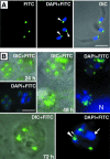
Fluorescence microscopy localization of Cp-mtHSP70 in homologous mitochondria. (A) Immunofluorescent antibody staining of purified C. parvum sporozoites with FITC-labeled anti-Cp-mtHSP70. The three-panel composite shows FITC, FITC plus DAPI double labeling, and a merged image with DIC. Note the single labeling (white arrowhead) in each of the three sporozoites at the posterior end and near the nucleus. Bar, 2.5 μm. (B) Immunofluorescent antibody staining of 24, 48, and 72 h C. parvum-infected HCT-8 cultured cells labeled with FITC anti-Cp-mtHSP70. Composite DIC and DAPI images are shown. Note the presence of DAPI nuclear staining in close proximity to FITC-labeled compartments. At 48 h, the host cell nucleus (N) is visible in the DAPI+FITC panel. At 72 h, globular nuclear material is indicated by an arrow; FITC labeling is indicated by the large arrowhead, and FITC-satellite labeling is indicated by the small arrowhead. Bar, 2.5 μm.
FIG. 7.
FIG. 7.
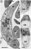
Transmission electron microscopy of C. parvum sporozoites showing the relict mitochondrion, its relationship to other organelles, and Cp-mtHSP70-specific immunogold localization within this organelle. (A) Longitudinal section of an epon-embedded freshly excysted sporozoite fixed in buffered 2% glutaraldehyde, with additional fixation in 2% osmium tetroxide and 0.5% uranyl acetate. This fixation clearly shows the organelles and their membranes. The ribosome-studded mitochondrion (*) is between the nucleus (N) and the crystalloid body (CB). The apical organelles shown include the micronemes (M) and dense granules (D) for entry into host enterocytes, as well as the plant-type storage granule amylopectin (A). Bar = 0.5 μm. (B) A series of three representative LR White-embedded sporozoites showing two, six, and three Cp-mtHSP70-specific immunogold particles labeling the relict mitochondrion (*). The localization of the compartment is identical to that seen in the osmium-fixed, epon-embedded sporozoites (Fig. 7A), i.e., the double-membrane-bounded organelle wrapped in ribosomes posterior to the nucleus in close apposition to the crystalloid body and previously identified as the C. parvum relict mitochondrion (40). LR White-embedded sporozoites were fixed in EM-grade methanol-free formaldehyde, with additional fixation in 4% formaldehyde-0.1% EM-grade glutaraldehyde. This fixative does not clearly reveal organellar membranes but is excellent for localization of immunogold antibody particles. Bar, 0.2 μm. Note the identical localization to this organelle using immunofluorescence (see Fig. 6A and B).

References

    1. Abe, Y., T. Shodai, T. Muto, K. Mihara, H. Torii, S. Nishikawa, T. Endo, and D. Kohda. 2000. Structural basis of presequence recognition by the mitochondrial protein import receptor Tom20. Cell 100:551-560. - PubMed
    1. Abrahamsen, M. S., and A. A. Schroeder. 1999. Characterization of intracellular Cryptosporidium parvum gene expression. Mol. Biochem. Parasitol. 104:141-146. - PubMed
    1. Altschul, S. F., T. L. Madden, A. A. Schaffer, J. Zhang, Z. Zhang, W. Miller, and D. J. Lipman. 1997. Gapped BLAST and PSI-BLAST: a new generation of protein database search programs. Nucleic Acids Res. 25:3389-3402. - PMC - PubMed
    1. Alvarez-Pellitero, P., and A. Sitja-Bobadilla. 2002. Cryptosporidium molnari n. sp. (Apicomplexa: Cryptosporidiidae) infecting two marine fish species, Sparus aurata L. and Dicentrarchus labrax L. Int. J. Parasitol. 32:1007-1021. - PubMed
    1. Arisue, N., L. B. Sánchez, L. M. Weiss, M. Müller, and T. Hashimoto. 2002. Mitochondrial-type hsp70 genes of the amitochondriate protists, Giardia intestinalis, Entamoeba histolytica and two microsporidians. Parasitol. Int. 51:9-16. - PMC - PubMed

Publication types

MeSH terms