. 2004 Mar 15;18(6):641-59.
doi: 10.1101/gad.1185804.

Palmitoylation is required for the production of a soluble multimeric Hedgehog protein complex and long-range signaling in vertebrates

Affiliations

Palmitoylation is required for the production of a soluble multimeric Hedgehog protein complex and long-range signaling in vertebrates

Miao-Hsueh Chen et al. Genes Dev. .

Abstract

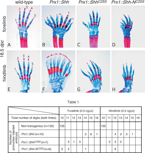
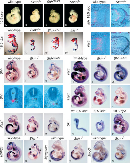
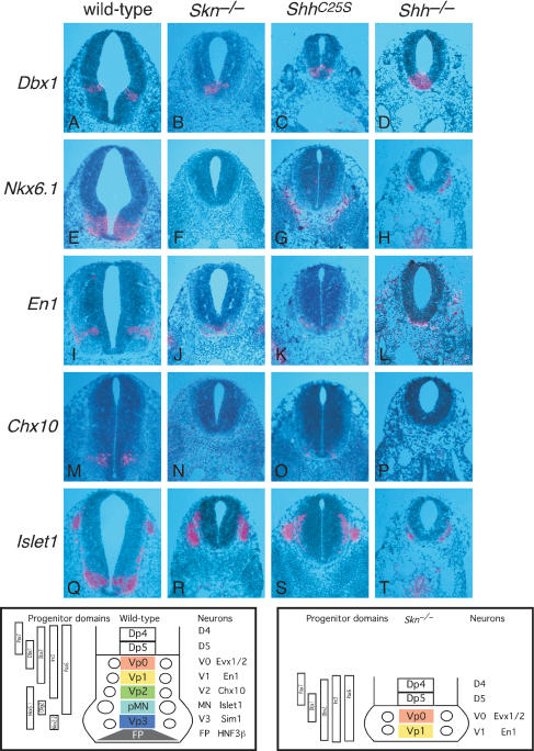
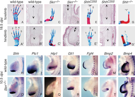
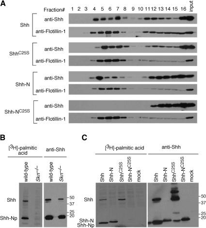
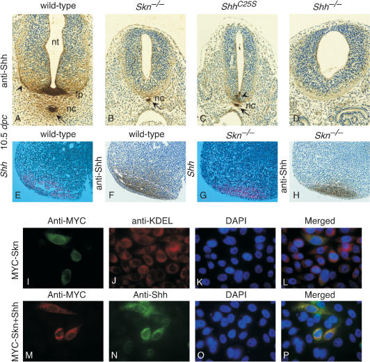
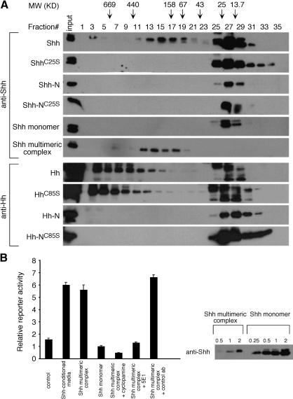
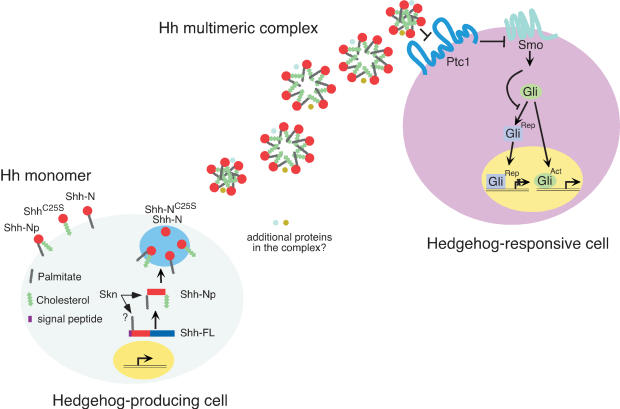
Hedgehog (Hh) signaling plays a major role in multiple aspects of embryonic development. A key issue in Hh signaling is to elucidate the molecular mechanism by which a Hh protein morphogen gradient is formed despite its membrane association. In this study, we used a combination of genetic, cellular, and biochemical approaches to address the role of lipid modifications in long-range vertebrate Hh signaling. Our molecular analysis of knockout mice deficient in Skn, the murine homolog of the Drosophila ski gene, which catalyzes Hh palmitoylation, and gene-targeted mice producing a nonpalmitoylated form of Shh indicates that Hh palmitoylation is essential for its activity as well as the generation of a protein gradient in the developing embryos. Furthermore, our biochemical data show that Hh lipid modifications are required for producing a soluble multimeric protein complex, which constitutes the major active component for Hh signaling. These results suggest that soluble Hh multimeric complex travels in the morphogenetic field to activate Hh signaling in distant Hh-responsive cells.


Figures

Figure 1.
Figure 1.
The activity of Hedgehog proteins with differential lipid modification in transgenic animals. Fore- and hindlimb skeleton (stained with Alcian blue [cartilage] and Alizarin red [calcified tissue]) of 18.5-dpc wild-type (A,E) and transgenic mice expressing Shh (B,F), ShhC25S (C,G), and Shh-NC25S (D,H) in the limb mesenchyme under the Prx1 promoter/enhancer. Views are all dorsal. The axis for orientation of the specimens is posterior on the right, anterior on the left, proximal downward, and distal upward. Table 1 summarizes the distributions of digit duplication in both fore- and hindlimbs of nontransgenic and transgenic animals expressing various forms of Shh proteins in the limb. Digits from both left and right limbs were counted, with 10 digits in nontransgenic wild-type mice. Compared with the hindlimbs, the forelimbs are more sensitive in their response to varying activities of Shh. Although the integration site of the transgene could potentially affect its expression levels, a general trend of decreasing activity from Shh to Shh-NC25S could be obtained by examining multiple transgenic animals. The DNAs were quantified and adjusted to 0.5 ng/μL for pronuclear injection.
Figure 2.
Figure 2.
Skn and ShhC25S mutants exhibit multiple defects due to reduced Hh signaling. (A-D) External morphology of wild-type, Skn-/-, ShhC25S, and Shh-/- embryos at 10.5 dpc. All views are lateral. (A′-D′) Skeleton of wild-type, Skn-/-, ShhC25S, and Ihh-/- 18.5-dpc embryos stained with Alcian blue and Alizarin red. All views are lateral. Embryos in A-D and A′-D′ were photographed at the same magnification, respectively. (E-V,H′-J′) Whole-mount in situ hybridization, using digoxigenin-labeled riboprobes on wild-type, Skn-/-, and ShhC25S embryos at 10.5 dpc with the exceptions of N (8.5 dpc) and O (9.5 dpc). All views are lateral. Expression of Hip1 is known to be completely dependent on Hh signaling (Chuang and McMahon 1999), whereas Ptc1 expression is initially Hh independent but is strongly up-regulated upon Hh signal transduction (Goodrich et al. 1996). Arrows in R point to residual HNF3β expression in the more rostral and caudal part of the ventral midline of the neural tube. Expression of Myogenin and MyoD (Tajbakhsh et al. 1997) in Skn-/- mutants cannot be readily distinguished from that in wild-type embryos. Embryos in E-G; H-J; H′-J′; K-M; Q and R; S and T; and U and V were photographed at the same magnification, respectively. (E′-G′) Isotopic in situ hybridization using 33P-UTP-labeled riboprobes (pink) on paraffin sections of wild-type, Skn-/-, and ShhC25S 10.5-dpc embryos at the forelimb/heart level. (E″,F″,H″,I″) Isotopic in situ hybridization using 33P-UTP-labeled riboprobes (pink) on paraffin sections of proximal tibia of wild-type and femur of Skn-/- 18.5-dpc embryos. Expression of Ptc1 in the chondrocytes is regulated by Ihh and not Shh (St-Jacques et al. 1999). (Nt) Neural tube; (fp) floor plate; (nc) notochord.
Figure 3.
Figure 3.
Skn and ShhC25S mutants exhibit defective short- and long-range signaling in the neural tube. (A-T) Isotopic in situ hybridization using 33P-UTP-labeled riboprobes (pink) on paraffin sections of wild-type, Skn-/-, ShhC25S, and Shh-/- 10.5-dpc embryos at the forelimb/heart level. The bottom left panel shows a schematic diagram of the expression domains of transcription factors, a combination of which define five progenitor cell types (Vp0, Vp1, Vp2, pMN, and Vp3) in wild-type embryos. As a result, five distinct neuronal types (v0, v1, v2, MN, and v3) are generated, which could be identified by neuronal-specific markers. In Skn mutants (bottom right panel) as well as in ShhC25S mutants, reduced Hh signaling leads to loss of floor plate, v3, MN, and most v2 with concomitant changes in the progenitor domains. This phenotype is similar to that in Shh-/- mutants perhaps with the exception of v2, a greater proportion of which is present in Skn and ShhC25S mutants. Similarly, the ShhC25S phenotype is slightly less severe than that in Skn mutants judged by residual v2. Expression of Islet1 in R-T represents neural crest-derived ganglia.
Figure 4.
Figure 4.
Skn and ShhC25S mutants have defective long-range Hedgehog signaling in the limb. (A,A′,C,C′,E,E′,G,G′) Fore- and hindlimb skeletons of wild-type, Skn-/-, ShhC25S, and Shh-/- 18.5-dpc embryos stained with Alcian blue and Alizarin red. All views are dorsal. The axis for orientation of the specimens is posterior on the right, anterior on the left, proximal downward, and distal upward. (B,B′,D,D′,F,F′) Hematoxylin-and-eosin-stained sections through the distal ulnar and tibia of wild-type, Skn-/-, ShhC25S, and Shh-/- 18.5-dpc embryos. Arrows indicate immature chondrocytes. (H-U) Whole-mount in situ hybridization, using digoxigenin-labeled riboprobes on wild-type and Skn-/- limbs at 10.5 dpc. Bmp4 expression in Skn-/- limbs cannot be distinguished from that in wild-type limbs (Jones et al. 1991).
Figure 6.
Figure 6.
The effects of lipid modification on Hh protein distribution in lipid rafts. (A) Western blots of fractions eluted from sucrose gradient probed with anti-Shh and anti-Flotillin-1 (a marker for lipid rafts) antibodies. Lysates from cells transfected with constructs encoding mouse Shh, ShhC25S, Shh-N, and Shh-NC25S, respectively, were processed for sucrose gradient fractionation as described in Materials and Methods. Fractions 4-7 represent lipid raft-containing fractions as indicated by the presence of raft-resident protein Flotillin-1 and caveolin (data not shown). Although Shh, ShhC25S, and Shh-N proteins could be detected in the lipid raft-containing fractions, no Shh-NC25S protein was present in these fractions. (B, left) Autoradiograph of immunoprecipitates of lysates separated on an SDS-PAGE. Lysates were derived from either wild-type or Skn-/- primary embryonic fibroblasts (MEFs) transfected with an expression construct encoding mouse Shh and grown in [3H]-palmitic acid-containing media. (Right) After exposure to film, the same membrane was processed for Western blotting probed with anti-Shh antibodies. Both unprocessed (Shh) and processed (ShhNp) Hh proteins were detected in wild-type and Skn-/- MEFs by Western blotting but only in wild-type MEFs could [3H]-palmitic acid-labeled Hh proteins be detected. (C, left) Autoradiograph of immunoprecipitates of lysates separated on an SDS-PAGE. Lysates were derived from HEK293 cells transfected with expression constructs encoding mouse Shh, ShhC25S, Shh-N, and Shh-NC25S, respectively, and grown in [3H]-palmitic acid-containing media. (Right) Similarly, after exposure to film, the same membrane was processed for Western blotting probed with anti-Shh antibodies. No [3H]-palmitic acid-labeled N-terminal fragment of Shh could be detected in lysates of cells transfected with ShhC25S or Shh-NC25S. Both full-length and processed Shh were labeled by [3H]-palmitic acid, as was Shh-N, indicating that cleavage and cholesterylation of Shh is not an absolute prerequisite for palmitoylation. The proportion of palmitoylated Shh-N remains to be determined. Numbers on the right indicate locations of protein size standards.
Figure 5.
Figure 5.
Shh protein is mainly restricted to its sites of synthesis in Skn and ShhC25S mutants. (A-D) Cross-sections of wild-type, Skn-/-, ShhC25S, and Shh-/- embryos at 10.5 dpc at the forelimb/heart level stained with anti-Shh antibodies. In the wild-type section, Shh immunoreactivity (brown) is strong in the notochord and floor plate, and it extends out bidirectionally in a graded fashion (arrows and arrowheads). Similar patterns of Shh immunoreactivity extending from the notochord were observed on embryo sections where the floor plate has not yet been induced (Gritli-Linde et al. 2001), suggesting that Shh immunoreactivity in the neural tube is derived from both the notochord and floor plate. In sections of Skn-/- embryos, Shh immunoreactivity is mainly detected in the notochord (arrow), and no obvious extended staining is present. Similarly, Shh immunoreactivity is mainly detected in the notochord (arrow) of ShhC25S embryos. In this section (C), residual Shh immunoreactivity could be detected in the ventral midline of the neural tube (arrowhead), but no obvious extension of staining to the neural tube was noted. This is due to a more posterior extension (to the forelimb level) of residual Shh mRNA expression in the ventral midline of the ShhC25S neural tube at the more rostral levels (data not shown) than that in Skn mutants (Fig. 2R). Immunostaining was performed on multiple sections and on sections where Shh mRNA (Fig. 2E′-G′) or protein expression level in the notochord of Skn-/- and ShhC25S mutants was comparable to that of wild-type; lack of spreading of Shh immunoreactivity was still observed. No Shh immunoreactivity is detected in the notochord or neural tube of Shh-/- embryos, although Shh immunoreactivity is detected in the gut of Shh-/- embryos, which represents cross reactivity to Ihh epitopes (data not shown). (E,G) Isotopic in situ hybridization using 33P-UTP-labeled riboprobes (pink) on paraffin sections of wild-type and Skn-/- limbs at 10.5 dpc. F and H are adjacent sections to E and G and were stained with anti-Shh antibodies. The dotted pink lines in F and H represent the approximate domains of Shh mRNA expression as determined in E and G. Multiple limbs were sectioned, and in situ hybridization and immunostaining were performed on all sections from the limb. (I-L) CHO cells transiently transfected with an expression construct encoding a MYC-tagged Skn and stained with anti-MYC and anti-KDEL antibodies (ER marker). L is a merged image of I-K. (M-P) CHO cells cotransfected with expression constructs encoding MYC-SKN and Shh and stained with anti-MYC and anti-Shh antibodies. P is a merged image of M-O. Similar results were obtained by using HeLa cells (data not shown). (nt) Neural tube; (fp) floor plate; (nc) notochord.
Figure 7.
Figure 7.
The effects of lipid modification on the generation of a soluble Hedgehog multimeric complex and protein activities. (A) Western blots of fractions eluted from gel filtration chromatography probed with anti-Shh antibodies. Supernatants collected from cells transfected with constructs encoding mouse Shh, ShhC25S, Shh-N, and Shh-NC25S, respectively, were processed for gel filtration chromatography as described in Materials and Methods. The fractions in which the molecular weight (MW) standards elute are indicated by arrows. The ratio between Shh multimeric complex and monomer varies between cell lines used in this study. The fractions corresponding to the multimeric and monomeric Shh, respectively, were subjected to a second round of gel filtration chromatography, and the fractions eluted were analyzed for the presence of Shh by Western blotting (labeled Shh monomer and Shh multimeric complex in the figure). Similar experiments were performed by using constructs encoding Drosophila Hh, HhC85S, Hh-N, and Hh-NC85S in S2 cells. Whether the high-MW protein complex that contains Drosophila Hh is biologically active or physiologically relevant remains to be determined. (B) Activity assays using the Shh-LIGHT2 reporter cell line. Shh multimeric complex exhibits greater activities in inducing Hh-response than Shh monomer as indicated by the luciferase reporter assay. The amount of protein used in this assay is shown on Western blots, in which the number 1 indicates the amount used in this assay, and the other numbers represent fractions or multiplications of that amount. The activity of Shh multimeric complex in this assay can be blocked by adding either cyclopamine or Hh-neutralizing antibodies (5E1) but not by a control antibody. Unfractionated conditioned media from HEK293T cells transfected with Shh also activated the Hh reporter and Shh-conditioned media contains at least fivefold more protein than Shh multimeric complex used in a similar assay.
Figure 8.
Figure 8.
A model of differential Hedgehog protein lipid modification and signaling. A schematic diagram of Hh protein biogenesis and transport. In Hh-producing cells, the Hh precursor molecule (Hh-FL) undergoes autoproteolytic cleavage to generate an N-terminal fragment, the mature form of which (denoted as Hh-Np; p indicates processed) becomes membrane-anchored likely due to the addition of a cholesterol group to the C terminus and a palmitoyl moiety to its N terminus catalyzed by Skn. Proteolytic processing precedes cholesterol modification but may not be a prerequisite for palmitoylation. The destiny of the C-terminal fragment after cleavage is not known. Hh-Np could self-associate spontaneously or in an active process that involves other proteins to generate a multimeric complex (depicted as a mixture of Hh hexamer, heptamer and octamer in this diagram although the actual number of Hh molecules in the multimeric complex may be variable), which is soluble and can be detected in the extracellular environment. Additional proteins may also exist in the Hh multimeric protein complex. The location of Hh multimeric complex formation is not known. The sequence and relationship between proteolytic cleavage, lipid modifications, multimer formation, and trafficking need to be further investigated. Hh multimeric complex is likely the vehicle to be transported to Hh-responsive cells to achieve long-range signaling, although the mechanism by which transport is achieved is unknown. Shh multimeric complex is more active than Shh monomer, and this could potentially be due to differences in affinity for the Hh receptor, Ptc1, or events downstream of ligand binding such as receptor oligomerization or modification. Hh binding to Ptc1 relieves Smo inhibition to allow Smo to transduce the Hh signal. As a result, generation of Gli repressor (Rep) is suppressed, whereas Gli activator (Act) is capable of activating Hh targets in the nucleus. When ShhC25S, Shh-N, and Shh-NC25 are expressed in Hh-producing cells under experimental conditions, a significant fraction of Shh-N still undergoes palmitoylation and both Shh-N and ShhC25S reach lipid rafts and can be detected in the extracellular environment in monomeric form. Whether interconversion occurs between Shh multimeric complex and monomer as well as between various Shh monomers in vivo is not known.

References

    1. Amanai K. and Jiang, J. 2001. Distinct roles of Central missing and Dispatched in sending the Hedgehog signal. Development 128: 5119-5127. - PubMed
    1. Ang S.L., Wierda, A., Wong, D., Stevens, K.A., Cascio, S., Rossant, J., and Zaret, K.S. 1993. The formation and maintenance of the definitive endoderm lineage in the mouse: Involvement of HNF3/forkhead proteins. Development 119: 1301-1315. - PubMed
    1. Bellaiche Y., The, I., and Perrimon, N. 1998. Tout-velu is a Drosophila homologue of the putative tumour suppressor EXT-1 and is needed for Hh diffusion. Nature 394: 85-88. - PubMed
    1. Brand A.H. and Perrimon, N. 1993. Targeted gene expression as a means of altering cell fates and generating dominant phenotypes. Development 118: 401-415. - PubMed
    1. Briscoe J. and Ericson, J. 1999. The specification of neuronal identity by graded Sonic Hedgehog signalling. Semin. Cell. Dev. Biol. 10: 353-362. - PubMed

Publication types