. 2004 Mar 15;18(6):715-27.
doi: 10.1101/gad.289504.

Cell cycle-dependent dynamic localization of a bacterial response regulator with a novel di-guanylate cyclase output domain

Affiliations

Cell cycle-dependent dynamic localization of a bacterial response regulator with a novel di-guanylate cyclase output domain

Ralf Paul et al. Genes Dev. .

Abstract

Pole development is coordinated with the Caulobacter crescentus cell cycle by two-component signaling proteins. We show that an unusual response regulator, PleD, is required for polar differentiation and is sequestered to the cell pole only when it is activated by phosphorylation. Dynamic localization of PleD to the cell pole provides a mechanism to temporally and spatially control the signaling output of PleD during development. Targeting of PleD to the cell pole is coupled to the activation of a C-terminal guanylate cyclase domain, which catalyzes the synthesis of cyclic di-guanosine monophosphate. We propose that the local action of this novel-type guanylate cyclase might constitute a general regulatory principle in bacterial growth and development.


Figures

Figure 1.
Figure 1.
Dynamic localization of the PleC and DivJ sensor protein kinases during the C. crescentus cell cycle. The positioning of PleC (circle) and DivJ (rectangle; Wheeler and Shapiro 1999) during the cell cycle are indicated. Polar organelles and cell cycle stages are specified.
Figure 2.
Figure 2.
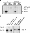
In vitro phosphotransfer between the protein kinases DivJ and PleC and the response regulator PleD. DivJ and PleC autophosphorylation in the presence of [γ-32P]ATP and subsequent phosphotransfer to PleD are shown. The bands corresponding to the phosphorylated proteins are marked on the side. (A) Assays contained 0.5 and 5 μg of the soluble kinase fragments and 37.5 μg GST-PleD as indicated. (B) Assays contained 12.5 μg DivJ′, 20 μg PleC′, and 50 μg PleD-H6 and PleDD53NH6, respectively, as indicated. In this experiment, DivJ′ and PleC′ were preincubated with [γ-32P]ATP for 15 min before PleD or PleDD53N were added to the reaction mix for an additional 5 min (PleC′) or 10 min (DivJ′), respectively.
Figure 3.
Figure 3.
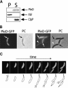
PleD is a soluble protein that localizes to the stalked pole of C. crescentus cells. (A) Immunoblot analysis of fractionated cell extracts of C. crescentus CB15N wild-type with anti-PleD, anti-FliF, and anti-ClpP antibodies. Cells of a logarithmically growing culture were lysed and soluble, and insoluble fractions were separated as indicated in Materials and Methods. Staining of the membrane-integral FliF and the soluble ClpP protein demonstrates the quality of the cell fractionation. (P) Insoluble pellet fraction; (S) soluble fraction. (B) PleD specifically localizes to the stalked pole. Phase contrast (PC) and fluorescent images of wild-type CB15N strains producing PleD-GFP from a low-copy number plasmid. The arrows indicate the polar foci of PleD-GFP in the fluorescent images and the stalk structures visible by phase contrast. (C) PleD dynamically localizes to the stalked pole during the C. crescentus cell cycle. Representative time-lapse experiment on C. crescentus wild-type cells producing PleD-GFP from a low-copy number plasmid. Fluorescent images (top) and a schematic representation of the cell cycle-dependent localization of PleD-GFP (bottom) are shown. In young swarmer cells, PleD-GFP is uniformly distributed in the cytoplasm. As the cells progress through the cell cycle and differentiate into stalked cells, PleD-GFP accumulates at the old flagellated and emerging stalked pole coinciding with flagellar ejection and stalk formation. PleD-GFP remains at the stalked pole throughout the cell cycle and is randomly dispersed in the newly formed swarmer cell. Only when the swarmer cell differentiates into a stalked cell again does PleD-GFP localize to the pole.
Figure 4.
Figure 4.
Dynamic localization of PleD to the stalked pole requires its activation by phosphorylation and is dependent on the polar kinases DivJ and PleC. (A) A nonphosphorylatable PleD mutant is impaired in polar localization. Phase contrast (PC) and corresponding fluorescent image of wild-type strain CB15N producing PleDD53N-GFP from a low-copy number plasmid. Bar: left panel, 2 μm. (B) DivJ and PleC are required for the polar positioning of wild-type PleD-GFP. Phase contrast (PC) and corresponding fluorescent images of wild-type and divJ pleC mutant strain producing PleD-GFP from a low-copy number plasmid. (C) DivJ and PleC are not required for localization of the constitutive mutant PleD*D53N-GFP. Phase contrast (PC) and corresponding fluorescent images of wild-type and divJ pleC mutant strain producing PleD*D53N-GFP from a low-copy number plasmid. (D) An active GGDEF output domain is not required for localization of PleD. Phase contrast (PC) and corresponding fluorescent image of a pleD mutant strain producing PleDGG368DE-GFP from a low-copy number plasmid. Filled arrows point to stalked poles in the phase contrast images (black) and to polar PleD-GFP foci in the fluorescent images (white). Open arrows point to nonpolar PleD-GFP foci.
Figure 5.
Figure 5.
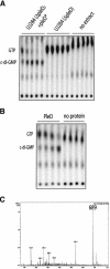
PleD is a di-guanylate cyclase. (A) Cyclic-di-GMP is produced by C. crescentus cell extracts. The soluble fraction of total cell extracts of CB15N ΔpleD (UJ284) or CB15N ΔpleD producing PleD* from a low-copy number plasmid were used to assay for di-guanylate cyclase activity. Control reactions without cell extracts are also shown. Samples were taken at 5, 10, 15, 20, and 30 min after addition of extracts and were analyzed on thin-layer chromatography plates. (B) Cyclic-di-GMP is produced by purified PleD protein. PleD-His6 (50 μg) was tested for di-guanylate cyclase activity, and the products of the enzymatic reactions were analyzed for 30, 45, 60, and 300 sec (lanes 1-4) after addition of purified PleD-His6, as indicated in A. (Lanes 5-8) Control reactions without PleD are also shown. (C) Analysis of products synthesized by PleD in vitro. The reaction products of PleD (200 μg) and GTP (1 mM) were separated by HPLC (cf. Fig. 6B). Peaks were collected and applied to mass spectrometry analysis. The fragmentation pattern shown corresponds to the main reaction product with a high-pressure liquid chromatography retention time of 6.73 min. The main peak had a molecular mass of 689 (theoretical molecular weight of c-di-GMP: 688.4). Reisolation and analysis of the substance with the molecular mass of 689 resulted in an identical fragmentation pattern.
Figure 6.
Figure 6.
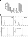
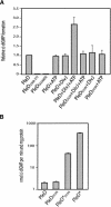
The PleD nucleotide cyclase activity is GTP-specific. (A) GTP, deoxyGTP, and c-di-GMP, but not ATP, specifically inhibit the PleD-dependent conversion of [32P]GTP into [32P]c-di-GMP. The relative c-di-GMP formation corresponds to the initial velocity determined for the enzymatic reactions. The reaction mixtures routinely contained 25 μg PleD and 100 μM GTP (see Materials and Methods) and were supplemented with nonlabeled nucleotides, as indicated below the graph. (B) PleD is a di-guanylate cyclase but lacks phosphodiesterase activity. High-pressure liquid chromatography analyses of GTP and c-di-GMP (left) and their reaction products with PleD (right) are indicated. The reaction mixtures contained 100 μg PleD and GTP and c-di-GMP at 200 μM each. Conversion of GTP into c-di-GMP by PleD is shown in the top panel by the appearance of a novel peak (arrow), which corresponds to chemically synthesized c-di-GMP (panels in second row). Incubation of PleD with chemically synthesized c-di-GMP for several hours did not lead to the cleavage or disappearance of the cyclic substance.
Figure 7.
Figure 7.
Activation of the PleD di-guanylate cyclase by phosphorylation and the requirement of an intact GGDEF output domain. (A) Influence of phosphorylation and mutations in the GGDEF domain on the PleD in vitro di-guanylate cyclase activity. The enzymatic reactions contained 100 μM GTP and were carried out with 25 μg of the following purified proteins: PleD, PleDD53N, PleDΔ368-372, and PleDGG368DE. DivJ (12.5 μg) and ATP (200 μM) were added where indicated. The relative c-di-GMP formation corresponds to the initial velocity measured for the enzymatic reactions. (B) Constitutive active forms of PleD mimic the activated state of the di-guanylate cyclase. The enzymatic reactions contained 200 μM GTP and were carried out with 12.5 μg of the following purified proteins: PleD, PleDD53N, PleD*D53N, and PleD*. The relative c-di-GMP formation corresponds to the initial velocity measured for the enzymatic reactions.
Figure 8.
Figure 8.
The GGDEF domain is coupled in a modular fashion with different sensory input or information transfer domains. The domain composition of GGDEF proteins listed in the SMART protein database (Schultz et al. 1998) is shown schematically. Recognized or putative signal sensing domains are indicated in rounded rectangles, the signal transfer domain (Rec) is indicated as a polygon, and the GGDEF domain is indicated as a rectangle. Connecting bars indicate the association of domains found in a single protein. The broken line symbolizes information transfer between sensor histidine kinases (e.g., DivJ) and the receiver domain of their cognate response regulator (e.g., PleD). (Rec) Receiver domain of response regulators; (MASE) membrane-associated sensor (MASE1 and MASE2; Nikolskaya et al. 2003); (PBPb) high-affinity periplasmic solute-binding protein of ABC-type amino acid transport system; (CHASE) cyclases/histidine kinases associated sensory exracellular (Anantharaman and Aravind 2001; Mougel and Zhulin 2001; Zhulin et al. 2003); (RXAG) permease component of ribose, xylose, arabinose, galactoside ABC transporter; (PAS/C:PAS) Drosophila period clock, aryl hydrocarbon receptor, and single-minded proteins (putative signaling domain; Ponting and Aravind 1997); (PAC) PAS C-terminal motif (Ponting and Aravind 1997); (MHYT) integral membrane sensor domain (Galperin et al. 1999); (SBP-bac) bacterial extracellular solute binding protein; (SHK) Sensor histidine kinase; (CACHE) signaling domain common to Ca2+ channels and chemotaxis receptors (Anantharaman and Aravind 2000); (GAF) cGMP-specific and -stimulated phosphodiesterases/adenylate cyclases (Anabaena)/FhlA (E. coli; Galperin et al. 2001); (HE) Hemerythrin, oxygen-binding protein (Stenkamp et al. 1978); (IQ) sequence motifs for calmodulin recognition (Rhoads and Friedberg 1997); (PL) phospholamban, small protein that regulates the affinity of the cardiac sarcoplasmic reticulum Ca2+-ATPase for calcium (Smith et al. 2001); (HAMP) Histidine kinases, adenylyl cyclases, methyl-accepting proteins, phosphatases (Aravind and Ponting 1999); (cNMP) cyclic nucleotide-monophosphate-binding domain.

Similar articles

Cited by

References

    1. Aldridge P. and Jenal, U. 1999. Cell cycle-dependent degradation of a flagellar motor component requires a novel-type response regulator. Mol. Microbiol. 32: 379-391. - PubMed
    1. Aldridge P., Paul, R., Goymer, P., Rainey, P., and Jenal, U. 2003. Role of the GGDEF regulator PleD in polar development of Caulobacter crescentus. Mol. Microbiol. 47: 1695-1708. - PubMed
    1. Amikam D. and Benziman, M. 1989. Cyclic diguanylic acid and cellulose synthesis in Agrobacterium tumefaciens. J. Bacteriol. 171: 6649-6655. - PMC - PubMed
    1. Anantharaman V. and Aravind, L. 2000. Cache—a signaling domain common to animal Ca2+-channel subunits and a class of prokaryotic chemotaxis receptors. Trends Biochem. Sci. 25: 535-537. - PubMed
    1. ____. 2001. The CHASE domain: A predicted ligand-binding module in plant cytokinin receptors and other eukaryotic and bacterial receptors. Trends Biochem. Sci. 26: 579-582. - PubMed

Publication types

MeSH terms